The Spinal Cord
Description
This section is from the book "A Manual Of Physiology", by Gerald F. Yeo. Also available from Amazon: Manual Of Physiology.
The Spinal Cord
The spinal cord, being the great bond of connection between the brain and the majority of the peripheral nerves, is necessarily a conducting apparatus of the very first importance, and from the quantity of nerve cells lying in its gray matter, it must also exercise the function of a governing organ, or nerve centre.
Spinal Cord As A Conductor. Anatomical Methods
Anatomical investigation shows that the spinal cord is not merely a collection or aggregation of the fibres that pass into it. In the first place, the spinal nerves, if bundled together, would be much larger than the cord, even at its thickest part; and secondly, it does not taper evenly toward its lower extremity, as it should were each succeeding pair of roots a direct loss of thickness. The question then arises, How are the fibres of the spinal nerves disposed of in the cord.

Fig. 242. Diagram illustrating the paths probably taken by the fibres of the nerve roots on entering the spinal cord. (Scha/er).
a.m./., Anterior median fissure; p.m.f., Posterior median fissure; c.c., Central canal; s.r., Substantia gelatinosa of Rolando; a a., Funiculi of anterior root of a nerve; p., Funiculus of posterior root of a nerve. By following the fibres 1, 2, 3, etc., their couise through the gray matter of the spinal cord may be traced.
The posterior roots of the spinal nerves (Fig. 242, p) pass through the white substance to reach the posterior gray column (SR), where they break up into twigs, some of which are distributed to neighboring parts of the gray network of fibrils, in which they are lost without their union with the cells being obvious or immediate, while others pass into the posterior white columns.
The fibres of the anterior roots, in irregular bundles (a, a), traverse the superficial white part of the cord on their way to reach the anterior gray columns, into the cells of which some can be directly traced; others enter thegray matter without joining the cells. The other processes from these cells pass into the fibrillar network which makes up the great mass of the gray substance, and are in communication with the distant parts of the cord above.
Anatomy may thus be said to leave us in the dark in regard to the paths traversed in the cord by the impulses on their way to or from the root of the spinal nerves.
Histology only teaches us that the gray and white matter of the cord consist of - (1i) innumerable fibrils and cells, and (2) medullated fibres variously connected with the different groups of cells and the roots of the spinal nerves.
Since ordinary histological research fails to show the complex connections of the fibres of the spinal cord, other methods have been resorted to in attempting to discover their course. Of these the two following have given good results:
Developmental Method
It was found that the medullary sheath of the fibres in different white tracts of the cord was perfected at different ages of an animal, and by tracing the course of the imperfectly developed fibres the functional relations of certain tracts could be arrived at.
Degenerative Method
It is well known that a degenerative process (sclerosis), easily recognized histologically, soon follows the section of nerve fibres. This change takes place first in those fibres situated at the side of the section toward which the impulses travel; i. e., at the peripheral end of an efferent and central end of an afferent fibre. By carefully tracing the course of the degenerated tracts after section or pathological lesion the function of the fibres and their connections with other parts of the nervous centres can be determined. 52
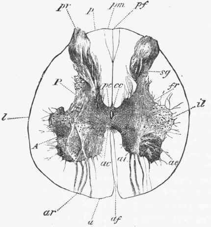
Fig. 243. Transverse section of the lumbar region of the spinal cord (for reference, see description under Fig. 245). (Sevan Lewis).
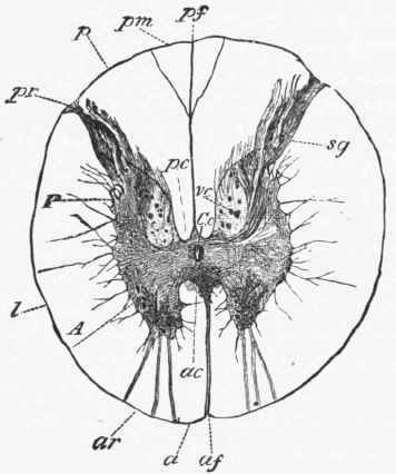
Fig. 244. Transverse section of the dorsal region of the spinal cord. (Bevan Lewis).
By these methods the following tracts of white fibres have been made out in the spinal cord:
1. The direct pyramidal tracts (Turck) (Fig. 246, T), each of which is continuous with the pyramid on the same side of the medulla oblongata. They lie on the inner aspect of the anterior white column, and form the immediate boundaries of the anterior median fissure. These tracts taper from above downward to nothing, and terminate about the middle of the dorsal region.

Fig. 245. Transverse section of the cervical region of the spinal cord.
A. Anterior gray column.
a. Anterior white column.
/. Lateral white column.
ac. Anterior commissure.
ar. Anterior roots.
of. Anterior median fissure.
tl. Intermedio-lateral gray column.
vc. Vesicular column of Clarke.
P. Posterior gray column.
p. Posterior white column.
pm. Posterior median column.
pc. Posterior commissure. • cc. Central canal.
pr. Posterior roots.
pf. Posterior median fissure.
ae and at. External and internal anterior vesicular columns. sg. Substantia gelatinosa.
The fibres leaving them appear to cross by the anterior commissure to reach the anterior gray column of the opposite side.
Continue to:
My Books

