Part 192. The Ferns - Plant Life-Histories. Part 2
Description
This section is from the book "Plants And Their Uses - An Introduction To Botany", by Frederick Leroy Sargent. Also available from Amazon: Plants And Their Uses; An Introduction To Botany.
Part 192. The Ferns - Plant Life-Histories. Part 2
We know that during the coal age many tree-ferns like the Pecopteris shown in Fig. 277, apparently near of kin to the adder-tongues, produced stout trunks bearing a crown of ample leaves nearly twenty meters above the ground.
At the present day tree-ferns such as the one shown in Fig. 360 abound in moist, warm regions, although the ferns most common in northern lands are more like the smaller ones shown in the same illustration. Thus it would appear that a certain amount of degeneration has attended the adaptation of ferns to the more stringent conditions of cold or dry climates. One of our best developed northern ferns is the Aspidium already studied (Fig. 170).
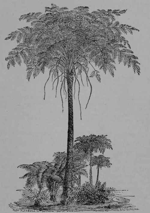
Fig. 360.-Tree-Ferns and Herbaceous Ferns. (Baillon.)
As shown in Figs. 361, 362, the spore in germinating produces first a row of cells, the terminal one of which soon divides in such a way as to produce a flat, heart-shaped thallus, which is rich in chlorophyll and sends out from the under side of the older part a number of pseudo-roots. By means of these the rear end is firmly attached to the earth while the lobed end slightly ascends. Finally on the lower surface appear archegonia near the tip, and male gametangia toward the base. The latter and their motile gametes are of the form shown in Fig. 363. The gametes, it will be noticed, are somewhat more highly developed than any found among the Bryophyta. That is to say, the spiral is larger and the flagella are more numerous. The archegonia, which are like the one shown in Fig. 364, differ but little from the others already studied. After fertilization the egg-cell divides into four (Fig. 365, A). The uppermost of these, by its further growth and division produces the foot (f) the function of which is to act temporarily as an haustorium for the embryo-sporophyte, and to push it out of the gametophyte and on to the earth. One of the lateral cells develops into the first root (w) while the opposite one becomes the growing point of the stem, and the lowest cell gives rise to the first leaf. A later stage in the development of these parts is shown in Fig. 365, B. Covering the growing tip of the root, somewhat as a thimble covers a finger tip, is a protective organ termed the root-cap. Such a thimble-like covering continually renewed by the meristem which it protects is characteristic of true roots. Root-hairs for absorption are soon developed. The leaf (Figs. 365, B, 362, B) soon differentiates into petiole and blade, and curves so as to drag the tender leaf-tip up out of the ground. An extreme curving of this nature performed by every branch of the developing leaves gives us the familiar crozier-like vernation characteristic of ferns. In the axis of the stem soon appears a central cylinder of prosenchyma which developing also in the root and the leaf serves as a channel for conducting solutions absorbed by the root to the green food-making parts of the leaf, and likewise dissolved nutrients from the leaves to the stem and the root where they may be used in growth or stored as a reserve. As the stem grows larger, and leaves and roots become more numerous, its central cylinder becomes a hollow cylindrical net-work of broad flat meshes (Fig. 366), giving off slender branches to the leaves and roots. When a leaf falls off it leaves a scar upon which one may see clearly traces of these slender branches which went into the petiole.
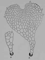
Fig. 361.-Male-fern (see also Fig. 170). a, b, germination of spores showing formation of young gametophytes (prothallia), 60/1. (Luerssen.)
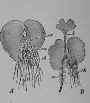
Fig. 362.-Male-fern. A, prothallus, lower side, showing archegonia (ar), antheridia (an), and pseudo-roots (rh), 6/1. B, same, after production of young sporophyte, showing first leaf (b) and first root (w). (Schenck.)
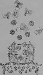
Fig. 363.-Fern Antheridium (Pteris sp., Polypody Family, Polypodiacece), 350/1. (Luerssen.)
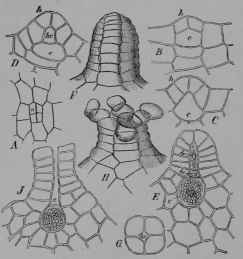
Fig. 364.-Fern Archegonium (Osmunda sp., Royal-fern Family, Osmundaceoe). A, first stage viewed from above, 80/1. B, same, cut vertically to show the central cell (c) from which the egg is formed, and the cells (h) which give rise to the neck, 120/1. C-E, older stages, showing canal cells (hc, bc). E, neck with mouth closed. G, same, top view. H, same, mouth open. J, same as E but with egg-cell (e) ready for fertilization. (Luerssen.)
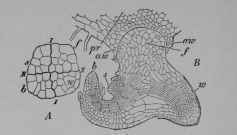
Fig. 365.-Fern Embryo (Pteris sp.). A, embryo removed from archegonium and cut vertically to show the first dividing wall (I, I) and the walls at right angles to this (II, II) whereby the fertilized egg-cell was divided into quadrants of which one (f) by further cell-division and growth becomes the foot, another (s) the stem, another (b) the first leaf, and another (w) the root. B, embryo still further developed but still attached to the prothallus (pr), cut vertically to show the foot (f) embedded in the archegonium (aw), the root (w) with its tip protected by a root-cap, the stem (s) and the incurved leaf (b). Magnified. (Hofmeister.)
Continue to:
My Books

