


3. Taenia Echinococcus
Description
This section is from the book "A Manual Of Pathology", by Joseph Coats, Lewis K. Sutherland. Also available from Amazon: A Manual Of Pathology.
3. Taenia Echinococcus
In the strobilus form this is a comparatively insignificant worm (Fig. 185). It inhabits the dog, and there are generally several individuals present at the same time. The total length of the worm is about an eighth of an inch, and it consists only of four segments, including that which carries the head. In the fully developed state the last segment exceeds in length the rest of the work together (see figure). The head is like that of the taenia solium in miniature, being very greatly less in size. It has a rostellum with thirty to forty hooklets, and four sucking discs. The last segment develops a large number of eggs, as many as 5000.
These eggs develop the usual embryoes with six spines, and if they find their way into the intestinal canal of man, the}' pass out into the tissues. Settling in some organ of the body, they show the most extraordinary powers of development, producing the condition commonly called Hydatids.
Hydatids occur in the form of large cysts, often of very complex arrangement, and they should be carefully distinguished from the cysticerci, which form small cysts not more than half an inch in size. The hydatids occur in the majority of cases in the liver. Neisser has collected no less than 986 cases of hydatids in man, and he gives the scale of frequency in the different organs as follows: - Liver, 451; lungs and pleura, 84; kidneys, 80; muscle and subcutaneous tissue (including the orbit), 72; brain, 68; spinal cord, 13; female organs of generation (including the mamma), 44; male organs, 6; pelvis, 36; organs of circulation, 29; spleen and bones, 28; eye, 3. Finsen found in Iceland that the lungs were affected only in 3 per cent, of the cases.
When the embryo reaches the liver or other resting-place, it soon develops into a cyst which at first is of slow growth. The membrane of the cyst is of considerable thickness, and consists of an external stratified cuticle .(the ectocyst), and an internal parenchymatous layer containing muscular fibre and a vascular system (the endocyst). Inside the original vesicle arise very frequently secondary vesicles, and inside these even tertiary ones, the successive vesicles being sometimes spoken of as daughter or grand-daughter vesicles. It sometimes happens that the secondary vesicles project outwards, and form a series of external vesicles which may separate from their mother and attain an independent development alongside of her. This latter form is particularly common in the domestic animals, and it is variously designated exogenous hydatids, and echinococcus scolecipariens or granulosus. Hydatids in bone usually assume the form of exogenous cysts (Targett). There is a third or alveolar form which has been met with in man, and always in the liver. In it the parasite develops a congeries of small vesicles, from the size of a grain of wheat to that of a pea. These are embedded in a gelatinous matrix and sometimes possess gelatinous contents. As the whole is surrounded by a firm fibrous capsule, the tumour is a somewhat solid one, and on section presents a peculiar alveolar appearance. This should be particularly borne in mind, as the condition has been frequently mistaken for a tumour, especially before Virchow demonstrated its true nature.

Fig. 185. - Adult taenia echinococcus. x 12. (Leuckart).
Except in the case of the alveolar form, the vesicles, both primary and secondary, enlarge very much and give rise to tumours of very large dimensions, so as sometimes to produce serious disturbance by their mere size. Those of the liver are usually the largest, and they may come to weigh as much as twelve, twenty, or even thirty pounds. The simple vesicles, in which no daughters develop, attain the size of an orange or a fist.
In all forms of hydatids the whole parasite is surrounded by a fibrous capsule, developed by the organ in which it has its seat. As the cysts enlarge, this also increases in size.
The formation of the heads of the worms differs in certain respects from that of the other taeniae. In the walls of the vesicles, either primary or secondary, are to be seen, when they are perfectly fresh, a number of small white points which have their seat in the internal wall. These are not the heads but the Brood-capsules or proligerous vesicles in the walls of which the echinococcus heads grow.
The heads begin (see Fig. 180, in which the development of heads in brood-capsules is shown) as projections outwards of the wall of the brood-capsule. The projection is hollow, and communicates with the interior of the brood-capsule. The head develops inside this projection, as in the case of the taenia solium, and very soon acquires the power of inverting itself. When it does so, it projects into the brood-capsule, so that in this respect the brood-capsule is not like the cyst of the cysticercus, as the scolex projects outwards from the latter. A single brood-capsule develops several heads, up to twelve, and they may be found either in the extended or inverted position. All the heads are contained in brood-capsules, but if after death or during removal the brood-capsule bursts, then an appearance may be produced as if the heads were attached to the wall of the cyst itself. If the capsule burst, its remains may gather round its stalk and the heads stand up from this as in Fig. 187, bb. Heads may also be found lying free if the capsules have burst. It is to be added that sometimes the vesicles remain barren, neither brood-capsules nor heads developing in them. These are the so-called Acephalocysts.
It is an interesting fact, that although the bursting of a hydatid cyst or the drawing off of its fluid generally causes the death of the parasite, yet that after rupture the parasite passing into the peritoneal cavity may live as minute vesicles, or grow so as to produce cysts as large as the fist.
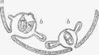
Fig. 186. - Diagrammatical illustration of development of echinococcus heads in brood-capsules, a, Wall of cyst; b. b, brood-capsules, with heads in various stages of development. (Leuckart).
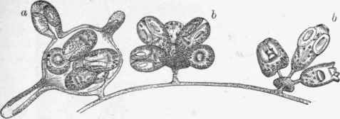
Fig. 187. - Brood-capsules in connection with walls of vesicle, a, A capsule in normal unruptured state; b, b, ruptured capsules, x 40. (Leuckart).
The heads which are formed in vast numbers inside the brood-capsules are exactly like those of the mature worm (see Fig. 187). They are very minute objects, measuring about 1/70th of an inch in long diameter, and just visible to the naked eye. They possess a proboscis with a ring of hooklets, and four suckers. They are also provided with a water-vascular system, and in their parenchyma abundant calcareous particles are to be found.
Continue to:


My Books

