


5. Tumours Of The Oesophagus
Description
This section is from the book "A Manual Of Pathology", by Joseph Coats, Lewis K. Sutherland. Also available from Amazon: A Manual Of Pathology.
5. Tumours Of The Oesophagus
Simple tissue tumours are rare. We meet with Lipomas and Fibromas, and the author has described a case of Myoma (Fig. 388) in which a tumour, 4 3/4 inches long and 2 inches in thickness, was attached by a comparatively narrow neck, and produced death by obstructing the tube. Polypoid tumours of a similar form are met with, having a fibrous structure.

Fig. 387. - Perforating ulcer in lower part of aesophagus. The ulcer penetrated the main bronchus of the left lung.
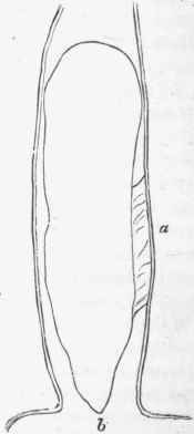
Fig. 388. - Polypoid myoma of oesophagus in section, and semi-diagrammatic. The tumour was attached by a band-like pedicle a. It caused considerable distension of the aesophagus, and obstructed the tube, b, cardiac orifice of stomach. Half the natural size.
Cancer of the aesophagus is by far the most important form of tumour. The cancer is nearly always in the form of flat-celled epithelioma, and in its histological details closely conforms to cancer of the lip. The masses of epithelial cells infiltrate the spaces in the underlying connective tissue, and the tumour also projects somewhat into the calibre of the tube. Here also there is a great tendency to ulceration, the mechanical action of the food in swallowing doubtless contributing to this result.
The tumour begins at a limited part of the mucous membrane, but it has a special tendency to extend round the tube in the form of a ring. There has been considerable discussion as to the most common situation of the tumour, and the result of the comparison of various statistics seems to be that the most frequent seat is the lower third. Scarcely less frequent than this is the middle third, and especially the place corresponding with the bifurcation of the trachea. In the upper third epithelioma is comparatively infrequent.
This form of tumour, when it surrounds the tube, frequently leads to Obstruction of the oesophagus. On examining the oesophagus after death the seat of the tumour is often indicated externally by a narrowing of the tube, which is also more rigid here than elsewhere. The infiltration of the walls of the oesophagus, by irritating the connective tissue, causes a chronic inflammation with the usual new-formation of connective tissue, which contracts and narrows the tube. Besides this, the mere rigidity of the infiltrated tube, preventing its dilatation when the morsel is being swallowed, may produce a virtual obstruction at the point concerned. The projection of the tumour into the calibre is another element, which tells especially in the earlier periods. But as ulceration occurs, this projection of the tumour usually becomes inconsiderable, and there may even be a temporary relief to the stricture by partial destruction of the tumour. The ulceration itself, however, by inducing still further cicatricial contraction, may ultimately confirm the obstruction.
The tumour sometimes extends from the oesophagus to neighbouring lymphatic glands, or to surrounding structures, and so we may have the trachea, bronchi, or lungs involved in it. Sometimes it extends to the diaphragm, pericardium, vertebrae, etc.
Literature
Knott, Path, of the aesoph. (with references to literature), 1878; Zenker, in Ziemssen's Encycl., vol. viii., 1878; Konig, in Billroth und Lueoke's Deut. Chirurg., xxxv., 1880; Klebs, (Diverticulum) Handb. d. path. Anat., i. (Esophagitis dissecans - Rosenberg, Centralbl. f. allg. Path., No. 18, 1892; Sela-vunos, Virch. Arch., cxxxiii., 1893, p. 250. Tuberculosis - Weichselbadm, Wien. med. Woch., 1884; Beck, Prag. med. Woch., 1884; Flexner, Johns Hopkins Hosp. Bull., iv., 1893. Syphilis - Weinlechner, Wien. med. Woch., 1880. Per-Joratiwj ulcer - Finlayson, Glasg. Med. Jour., xix., 1883, p. 313; Flower, Med. chir. trans., xxxvi., 1853; Hilton, Path, trans., vi., 1854; Part, ibid., viii.; Quincke, D. Arch. f. klin. Med., xxvi., 1879; Kehrer, ibid., xxxvi., 1885. Tumours - Virchow, Geschwulste, iii.; Robenbach, (Sarcoma) Berl. klin. Wochenschr., Sept. 20 and 27, 1875; Coats, (Myoma) Glasg. Med. Jour., iv., 1872, p. 201; Fagge, (do.) Path, trans., xxvi., 1875, p. 94.
Continue to:


My Books

