Anatomical Changes In Phthisis. Part 4
Description
This section is from the book "A Manual Of Pathology", by Joseph Coats, Lewis K. Sutherland. Also available from Amazon: A Manual Of Pathology.
Anatomical Changes In Phthisis. Part 4
The Bronchiectatic cavity is lined with a distinct membrane, and is usually directly continuous with a bronchus (see Fig. 369). It may exist in the midst of crepitating lung tissue, the complementary dilatation being in a part not affected by the tuberculosis.
Emphysema is a frequent accompaniment of the process. Wherever a piece of lung escapes the fibroid change, it is liable to emphysema on account of the shrinking in its neighbourhood. Hence in the midst of the shrunken tissue one often sees islands of lung tissue having a honeycombed appearance (see Figs. 371, b, and 373, b).
Another occasional result of the shrinking is a formation of Cyst-like cavities in the pleura, as shown in Fig. 373, a. The shrinking of the lung, dragging the adherent pleura with it, may cause spaces to form in the pleura, of even in the interlobular septa (as at c), these spaces being filled with serous fluid.
Pigmentation is a peculiarly prominent feature in fibroid phthisis. Even the initial lesion is characterized by the almost black colour of the nodules, and the indurated tissue has a slaty or blackish colour (slaty induration). Perhaps the explanation of this is that the carbonaceous pigment (see further on) is retained by the affected bronchi and not swept outwards by the cilia of the epithelium.
The caseous and fibroid forms of phthisis are in general distinguishable. They have certain points in common, chiefly in respect that each begins with a bronchitis of the finer tubes, and that each is characterized by the presence of tubercles. In the one form, however, the bronchial inflammation extends to the proper parenchyma of the lung, constituting a Lobular bronchopneumonia, whereas in the other it is more localized around the inflamed bronchus, constituting a tubercular Bronchitis and Peri bronchitis. There is the further distinction that in the one form caseous necrosis is characteristic, whereas in the other, while probably present in most cases, it is limited in extent and may be confined to the contents of the bronchi and the bronchial wall.
The difference is probably due to differences in the individual proclivities of the persons affected. In the fibroid form the disease is more chronic, the persons affected are as a general rule older, and it occurs more frequently in the male sex. All these facts point to the conclusion that in this form there is greater resistance on the part of the tissues to the morbid poison. This is confirmed by the resemblance which the process, in some respects, presents to that which is concerned in the healing of phthisis. It may be said that in the caseous form the tissues are directly killed by the progress of the disease, sometimes with great rapidity, whereas in the fibroid form there is a long struggle and very little palpable softening or destruction.
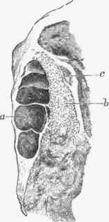
Fig. 373. - Fibroid phthisis. a, cysts in pleura from shrinking of lung; 6, emphysema; e, cyst in interlobular connective tissue. Natural size.
This being the case it may be inferred that the two forms are not absolutely distinguishable. They run into each other, and the caseous form may assume many of the characters of the fibroid, especially when it becomes very chronic or partial recovery takes place.
The Sputum in phthisis pulmonalis is variously composed. In the earlier stages the expectoration has the usual characters of that in catarrh, consisting of mucus, with more or less abundant leucocytes. In the sputum in this early stage are often found large epithelioid cells with one or more nuclei, such as we find in the lung alveoli in the catarrhal form of phthisis. These cells frequently present fatty degeneration. The sputum in phthisis often contains Elastic tissue from the breaking down of the lung. In very rapid cases we may find this by a simple examination of the sputum, but the search is often a difficult one, because the thick mucus and pus hold the pieces of lung tissue suspended and isolated. By Fenwick's method of digestion in soda solution, pieces of lung tissue, such as that shown in Fig. 368, will frequently be found in the deposit. This method is also applicable to the sputum in gangrene of the lungs. The Tubercle bacillus is usually to be found in the sputum, and is of great diagnostic value. The appearances are shown in Fig. 127, p. 304, and the method of examining the sputum is described at p. 360.
Continue to:
My Books

