Diseases Of The Arteries
Description
This section is from the book "A Manual Of Pathology", by Guthrie McConnell. Also available from Amazon: A Manual Of Pathology.
Diseases Of The Arteries
Endarteritis, or inflammation of the artery, usually results from the presence of foreign bodies, either infectious or sterile, within the vessel. It may be caused by organisms gaining entrance into the vasa vasorum. The intima is first involved; it becomes roughened, the endothelial cells become loosened, and there is usually an infiltration of round cells. The vasa vasorum are involved and the inflammatory process may extend to the media or the adventitia, and as a result a thrombus generally forms within the lumen. This may undergo organization with connective-tissue formation within the lumen of the vessel - thromboarteritis proliferans.
Periarteritis is an inflammatory condition around an artery, usually arising from injuries from without, or sometimes by extension from within. There is an infiltration of the adventitia, which becomes swollen and edematous. The media and intima become involved, there is desquamation of the endothelium with the formation of thrombi, usually infectious.
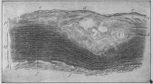
Fig. 136. - An Atheromatous Patch in the Abdominal Aorta which Has Not Yet Broken Through (Dmitrijeff).
J, Intima; M, media; A, adventitia; b, atheromatous necrotic focus in the intima and media; c, elastic fibers of the intima; d, elastic fibers which have persisted between the necrotic focus and the endothelial layer; e, thickened endothelium; /, infiltration of the media with small cells.
Arteriosclerosis, arterio-capillary fibrosis, or chronic arteritis, is a condition characterized by an increase in connective-tissue formation accompanied by degenerative changes. These may be circumscribed or diffuse. The fibrous formation occurs chiefly in the outer coats and is referred to as sclerosis, the degenerative processes involve the sub-intimal tissue and are spoken of as atheroma.
The large vessels such as the aorta, particularly the arch, are most commonly affected, but the arteries at the base of the brain and the splenic artery are frequently involved. Such an involved vessel, as the aorta, is usually dilated and the intima greatly roughened. The surface appears mottled on account of the varying changes from the normal yellowish streaks of atheroma, pale areas of sclerosis and calcification will alternate with reddish collections of fibrin.
In the circumscribed form, arteriosclerosis nodosa, numerous small oval or round yellowish-white areas are visible. These are but slightly elevated and vary in their consistency according to the structure. If there is much connective tissue, they may be very firm, almost cartilaginous; if degenerative changes have occurred, they will be soft.
As a result of the presence of these areas the elasticity of the vessel is interfered with, nutrition suffers, and connective tissue forms. Beneath the intima this new-formed fibrous tissue, which is usually dense, tends to undergo fatty degeneration, to become necrotic and to degenerate. In this way large or smaller cavities, filled with a softened semi-fluid substance consisting of disintegrated tissue, fat, and cholesterin crystals, develop. They are called atheromatous cysts and are covered by an imperfect layer of endothelium. This covering may break off, allow the contents to escape, leaving a cavity known as an atheromatous ulcer. The material found within these so-called cysts is composed of tissue that has undergone a fatty degeneration. Microscopically yellowish granules and droplets of fat as well as crystals of fatty acids are present. Instead of escaping, the cystic contents may become markedly infiltrated with lime, thus forming atheromatous plates.
Whenever an atheromatous ulcer has formed, the wall of the vessel at that point may be thinner and less elastic than normal. Eventually there may be a proliferation of the connective-tissue coat to give support to the weakened area. As the elasticity of the vessels is decreased there is consequent increase in blood-pressure with hypertrophy of the left ventricle. With the increased pressure there occur dilatations of the vessel at its weakened points, with the formation of aneurysms.
In the smaller vessels, particularly of the brain, rupture or apoplexy quite frequently occurs.
In diffuse arteritis or arterio-capillary fibrosis the smaller arteries and capillaries are the seat of fibrous tissue formation. Resulting from this, there may be more or less diminution of the lumen of the vessel, endarteritis deformans.
If the lumen is completely occluded, is known as endarteritis obliterans. In both cases there will be interference with the supply of nutrition and degenerative changes, chiefly hyaline, of varying degree consequent.
These lesions are very common in both syphilis and tuberculosis. In the first there is a cellular proliferation beginning in the adventitia but ultimately involving the intima.
Continue to:
My Books

