Life-Cycle Of Plasmodium Vivax (After Grassi And Schaudinn)
Description
This section is from the book "A Manual Of Pathology", by Guthrie McConnell. Also available from Amazon: A Manual Of Pathology.
Life-Cycle Of Plasmodium Vivax (After Grassi And Schaudinn)
The human cycle is above the transverse line, somewhat rearranged by Kisskalt and Hartmann. The cycle in the mosquito is beneath, 1 to 7, schizogony; 1, sporozoite; 2, entrance of the sporozoite; 3, 4, growth of the schizont; 5, 6, nuclear division of the schizont; 7, formation of the merozoites; 8, merozoites; 9a to 12a, growth of the macrogametocyte; 9b to 12b, growth of the microgametocyte; 13c to 17c, parthenogenesis of the macrogametocyte; 13a. 14a, maturation of the macrogamete; 13b, 146, growth of the microgamete; 15b, microgamete; 16, fructification; 17, oöki-net; 18 to 20, entrance of the oökinet into the stomach-wall of the mosquito; 20 to 25, sporogony; 22, 23, nuclear multiplication in the sporont; 24, 25, formation of the sporozoites; 26, passage of the sporozoites to the salivary gland; 27, salivary gland of the mosquito with sporozoites. (Magnification, 1 to 17c, 1200 to 1; 18 to 27c, 600 to 1).
(Park and Williams' "Pathogenic Micro-organisms.")
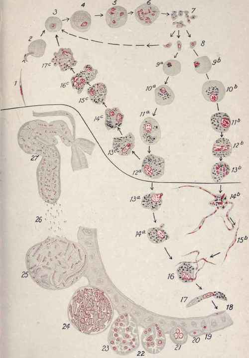
PLATE III. Life-cycle of Plasmodium Vivax (after Grassi and Schaudinn).
When the blasts gain entrance into the blood, they attack the red corpuscles and give rise to malaria.
In this case the mosquito is the definitive host of the parasite, man the intermediate.
The cycle of development in the mosquito varies according to the type of malarial organism - about three weeks in the quartan, two weeks in the tertian, and twelve days in the estivo-autumnal.
The following is a description of the Anopheles mosquito. Palpi in both sexes, nearly as long as the proboscis, 4 jointed in females, 3 in male. Is a constricted basal joint in each. Palpi are straight and parallel with proboscis except when female is biting, then they diverge.
Nucha has scaly posterior cornu, abdomen hairy on both surfaces, not scaly. Legs long and end in simple dentate claws. Wings spotted, and these spots when magnified are seen to be made up of black squamae on brownish wing.
Length of female 7.5 to 9 mm., including proboscis; male smaller and does not bite.
When resting on a perpendicular wall the Anopheles extends its body at right angles unless it is filled with blood, the Culex holds its body parallel.
Yellow fever is an infectious disease, probably caused by some protozoön which is carried by a mosquito, the Aedes (Stegymia) calopus.
The female mosquito is from 3.5 to 5 mm. long, head clothed with flat scales, black and gray on each side. A white patch in the middle in front, extending back to the neck. A white patch on each side and thin white borders to the eyes. Antennae blackish with narrow pale bands. Last joint of palpi white on inner side. Thorax dark brown, ornamented with white curved band on each side of the back and white spot on each side in front.
Abdomen dark with basal bands of white.
Fore- and mid-ungues toothed, hind ones not.
This mosquito may convey yellow fever to a non-immune as early as on the twelfth day after biting an infected person, and it may retain the power to do so as long as it lives.
This disease can also be transmitted by the hypodermic injection of blood drawn from a patient in the first, second, or third days of the disease. It cannot be communicated by fomites.
The infected agent can be passed through a filter that is impermeable to ordinary bacteria and is destroyed by a temperature of 550 C. maintained for ten minutes.
One attack usually renders a person immune.

Fig. 102. - Trypanosoma, showing Multiplication by Division (fromLaveran).
n, Nucleus; c, centrosome; m, undulating membrane; f, flagellum.
Continue to:
My Books

