The Haemameba Malariae
Description
This section is from the book "A Manual Of Pathology", by Guthrie McConnell. Also available from Amazon: A Manual Of Pathology.
The Haemameba Malariae
The Haemameba Malariae is a unicellular protozoan parasite that, during one cycle of its existence, lives in the blood and brings about a destruction of the erythrocytes. Its other cycle is carried on within the body of a mosquito, the Anopheles.
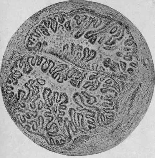
Fig. 101. - Coccidiosis of Rabbit's Liver (McFarland).
Section of one of the affected bile-ducts, showing the papillary outgrowths from the mucous membrane and the signs of inflammation in the surrounding tissue.
There are three varieties of the organism:
1. The quartan parasite, Hoemanieba malaria, attains its development in three days. It appears inside an erythrocyte as a small, unpigmented, irregular, hyaline body capable of slow ameboid movement. It grows gradually, and brownish or black granules appear within a few hours. The erythrocyte becomes gradually paler and is finally completely filled by the Plasmodium.
The pigment occupies the center of the cell, and later the parasite splits up into from six to twelve pear-like segments which, along with the pigment, escape into the circulation. Is the rosette form. The granules in this form are larger and darker than in the tertian, but not so numerous.
Double infection may occur, in which case there would be paroxysms for two days, then an intervening free day.
2. The tertian parasite, Hoemameba vivax, requires two days for its development. In its early stages it resembles the quartan parasite, but it eventually becomes larger. The tertian continues growing until it may be double the size of a red cell, usually but slightly larger. The pigment particles appear in about six hours, at first scattered, but finally collect in the center and are actively motile. The organism then divides into fifteen to twenty small, round, spore-like bodies, resembling a bunch of grapes. This form of parasite contains more granules than the quartan, but they are smaller and the red corpuscle is rapidly decolorized.
3. The oestivo-autumnal parasite, Hoemameba falciparum, is probably a tertian form. Is sometimes spoken of as malignant tertian.
The cycle of development lasts forty-eight hours. The organism is smaller than in the first two, but more active. When fully developed, it is about one-third the size of the red cell.
When at rest, it assumes the signet-ring form - a disc with a colored outline. The pigment develops within twenty-four hours in the form of a few coarse granules centrally located; is non-motile.
The organism finally becomes lobulated, rosette-shaped with the pigment in the center or toward the periphery, and divides into six to twelve little balls.
The development of this form seldom takes place in the peripheral blood, usually in the spleen, bone-marrow, and cerebral capillaries.
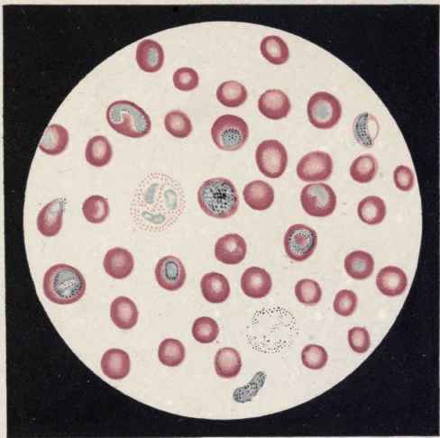
PLATE II. Malarial Parasites in Blood (Grawitz).
Numerous Pigmented Parasites, Spore Formation Of The Tertian Type, And Two Crescents
Table Showing Chief Differences Between the Species of Malarial Organisms (Genus HAemameba) Found in Man (Park and Williams). | |||||||||
Name of organism | Size of parasite up to segmentation (schizont) | Motion of young schizont in corpuscle | Time of appearance of melanin granules and their arrangement | Shape of segmenting parasite, number of segments, and site of segmentation | Asexual cycle complete in | Sexual forms | Incubation period | Effect on human host tissues | Remarks |
H. vivax (parasite of tertian fever). | 1 µ to slightly larger than normal red blood-cell (may occasionally be almost twice size). | Markedly active. | 6 hours. First scattered, then gathered in center. Finely granular. Actively dancing. | Irregular mulberry. 12-24 (average 16). Peripheral circulation. | hours. | Gametocytes spherical. No crescents. Male 2/3 size of female, which is 1 ½ times size of red blood-cell. No crescents. | About 14 days. | Pale, granular, slightly enlarged red-blood-cells. Finely granular pigment formed from metamorphosed hemoglobin. | Double infection may cause a paroxysm every day, thus giving clinically a quotidian type of fever. |
H. malariae (parasite of quartan fever). | 1 µ to little less than size of blood cell. | Not very active. | Within a few-hours. Collected in zone on periphery. Coarsely granular. Slight dancing. | Regular daisy shape. 6-10 (average 8). Peripheral circulation. | 72 hours. | Gametocytes spherical. Fewer than in vivax. About size of red blood-cell. No crescents. | About 3 weeks. | Red blood-cells may be slightly shrunken. | Double infection may cause a paro-oxysm every day, thus giving clinically a quotidian type of fever. |
H. falciparum (parasite of estivo-au-tumnal (pernicious) fever type). | Smaller than others, from very small to ½ diameter corpuscle. | Active, but slightly less so than tertian type. --------- | Within 24 hours. Small amount, 2-3 coarse granules usually central. Non-motile or sluggish. | More or less symmetrical daisy. 10-32 very small. Chiefly in bone marrow and viscera. | 24-48 hours. | Gametocytes crescentic, short and plump. ¾ size of red cell. | About 10 days. | Red blood-cells unstained, greenish, shriveled (crenated) and darkened; stained (Giemsa) salmon color. | |
The erythrocytes tend to shrivel and become dark. In this variety of infection are found the crescents of Laveran. They are oval or crescentic bodies that are pigmented in their center and have no ameboid motion, but are able to slowly alter their shape.
When fully developed are larger than a red cell. They are found partly in red corpuscles, or clinging to them, free in the blood and sometimes in leukocytes. Occur only in the severe forms of malaria; are probably malignant tertian parasites that have failed to sporulate.
When the organisms sporulate and are set free within the blood, the fever rises and the chill takes place. The escaped spores enter other red corpuscles, go through the same cycle, and a continuous reinfection occurs.
The foregoing cycle is the asexual one, schizogony, that takes place within the human body. It is now definitely settled that infection takes place by means of the bite of certain mosquitoes belonging to the genus Anopheles. Of mosquitoes, only the female bites.
This insect withdraws the malarial parasites from an infected individual; these undergo the sexual stage of development, sporogony, and are again transmitted to other people by the mosquito biting.
In the blood of an infected person two chief forms of parasite are found. One that is the smaller is round and contains numerous granules and is called the microgametocyte. From it flagella - microgametes - are given off. These flagella penetrate a larger spherical form that has a clear protoplasm, the macrogamete, which has formed from the macro gametocyte by the extrusion of small reduction nucleus; the former being the male, the latter the female, element. The impregnated parasite is called a zygote. These zygotes penetrate the stomach and become attached to the outer wall, where they grow much larger and are called sporocysts. They finally undergo division into secondary spheres, sporoblasts, which ultimately split up into very many small, spindle-like bodies known as sporozoites. These escape into the body cavity of the mosquito and the majority finally gain entrance into the salivary glands, from which they escape in the secretions.
Continue to:
My Books

