Thrombosis
Description
This section is from the book "A Manual Of Pathology", by Guthrie McConnell. Also available from Amazon: A Manual Of Pathology.
Thrombosis
Thrombosis is the coagulation of the blood within the vessels during life. It may depend upon changes within the blood, changes in the cardiovascular structures, and diminution of the velocity of the blood-flow.
The changes of the blood are those which tend to increase its coagulability. In the formation of a thrombus there is an action of the fibrin ferment or thrombin upon certain of the proteins in the blood-plasma. This ferment is not present in the normal circulating blood, but is produced after the blood is discharged from the vessels by the action of thrombokinase upon the thrombogen of the plasma in the presence 6i calcium salts. The thrombokinase is supposed to be liberated by the breaking down of leukocytes and blood-platelets. Certain chemical and physical substances - alcohol, ether, chloroform, heterologous blood-serum - when in the circulation may liberate fibrin ferments and thus cause thrombosis. The toxins of pneumonia, of diphtheria, and those resulting from extensive burns are especially active.
The lesions of the vessel walls are particularly important. Fibrin will be deposited upon the wall of the heart or bloodvessels whenever the nutrition of the endothelium of that wall is impaired. Diseases leading to the roughening of the endothelium, particularly arteriosclerosis, are important causes. Inflammation of neighboring structures may bring about changes within the intima. Ligation of a vessel causes an injury to the internal coat, and in that way predisposes to coagulation.
Diminution of the blood-flow may result not only from cardiac disturbances, but also from conditions causing a decrease in the lumen of the vessel. As the current slows, the leukocytes tend to adhere to the wall of the vessel, blood-plates make their appearance, and fibrin is deposited. The nutrition of the endothelium suffers, changes take place in the wall, and another factor in thrombosis then arises. The appearance of a thrombus depends upon the number of red corpuscles contained within it, and that rests upon the varying rapidity of the blood-current at the time of formation. It is generally made up of superimposed layers of fibrin. After a thrombus has formed there is always a tendency for it to extend up the vessel, against the current of the blood, and to involve successive branches.
If the blood were passing through the vessel with considerable velocity, the thrombus would be grayish-white in color, and on section would show well-marked lamination. This is called a white thrombus.
If the blood were moving less rapidly, varying numbers of red cells would be entangled in the fibrin and the color would be brown or grayish red, giving rise to a mixed thrombus.
If it is formed in a short time from blood that is barely moving, a red thrombus will result.
A true thrombus differs from a post-mortem clot within a vessel in that the latter is moister, is never adherent to the vessel wall, and never laminated. The clot may show also a division into pale, "chicken fat," and dark "currant jelly" portions as a result of the coagulation taking place after the heavier red corpuscles have sunk.
Thrombi
Thrombi may be classified according to their etiology as:
1. Infectious - those depending upon the entrance of bacteria into the circulation.
2. Mechanical - foreign bodies free from organisms. According to their period of formation as:
1. Primary or initial thrombi.
2. Secondary or consequential, depending upon a pre-existing thrombus and usually extending to the first collateral branch of the blood-vessel.
According to their morphology as:
1. Central, occluding, or obstructing - formed by the coagulation of the entire mass of blood contained within a certain portion of the vessel.
2. Parietal - when attached to the wall of the vessel, but not completely obstructing it.
3. Valvular - parietal thrombi that have become partially detached.
4. Channeled or tunneled - those in which there still exists a lumen through which the blood can pass. May be the result of secondary changes in old thrombi.
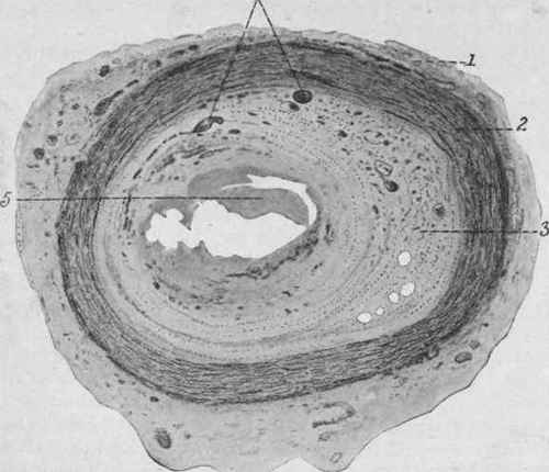
Fig. 3. - Organized and Partly Canalized Thrombus of the Brachial Artery. X 32 (Dürck).
1, Adventitia; 2, tunica media; 3, organized thrombus - i. e., replaced by connective tissue; 4, newly formed and in part dilated vessels within the thrombus; 5, disintegrated remains of the old thrombus.
5. Ball - thrombi that lie free within the cavities of the heart, usually in the auricles.
6. Polypoid - -ball thrombi with pedicles.
Continue to:
My Books

