Pathology. Origin Of Lesions
Description
This section is from the book "Early Detection And Diagnosis Of Cancer", by Walter E. O'Donnell. Also available from Amazon: Early Detection And Diagnosis Of Cancer.
Pathology. Origin Of Lesions
Most breast cancer is adenocarcinoma, which arises from the mammary duct epithelium.
The terminal ramifications of the mammary duct system are organized in clusters that form lobules. In its early stages breast cancer may be called intraductal carcinoma, with noninfiltrating (i.e., in situ) and infiltrating stages often delineated. When breast cancer originates in a lobule, it may be called lobular carcinoma.
Paget's disease of the breast is a form of duct carcinoma that first presents as an eczematoid lesion of the nipple.
Histologic Classification
Extensive classifications of breast cancer by histopathologic types have been proposed but need not concern the nonspecialist. Among the more frequent types, noninfiltrating mammary duct carcinoma, whether of the papillary or comedo variety, and infiltrating carcinoma of the medullary and colloid type tend to have a relatively more favorable prognosis than scirrhous carcinoma. Lymphosarcoma and the various stromal sarcomas have a better prognosis than the breast carcinomas in general.
Location Of Lesions
Most cancers of the breast arise in the upper outer quadrant (Fig. 46). A significant number are located in the central or subareolar area of the breast. Those breast cancers arising centrally and in the medial quadrants have a special significance therapeutically and prognostically since they are more apt to spread to the internal mammary nodes than are cancers arising in the outer quadrants.
Pathways Of Spread
Although breast cancer may first appear as a distant metastasis it tends to spread first through regional lymphatic pathways. The usual routes are the axillary nodes for lesions located laterally and the internal mammary nodes for those located centrally and medially.

Fig. 45. Anatomy of the breast.
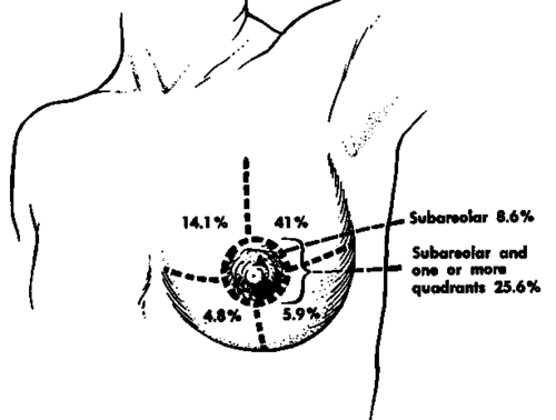
Fig. 46. Distribution of cancer of the breast. (Based on data from the Breast Service, Department of Surgery, Memorial Hospital for Cancer and Allied Diseases.).
In recording the extent of axillary node involvement, the pathologist may use the following node levels:
Level I All nodes below the inferior border of the peetoralis minor muscle
Level II All nodes lying beneath the peetoralis minor muscle
Level III All nodes above the superior border of the peetoralis minor muscle
Continue to:
My Books

