Chapter V. Conducting Tissue
Description
This section is from the "Histology of Medicinal Plants" book, by William Mansfield. Also see Amazon: Histology of Medicinal Plants.
Chapter V. Conducting Tissue
All cells of which the primary or secondary function is that of conduction are included under conducting tissue. It will be understood how important the conducting tissue is when the enormous quantity of water absorbed by a plant during a growing season is considered. It will then be realized that the conducting system must be highly developed in order to transport this water from one organ to another, and, in fact, to all the cells of the plant. Special attention must be given to the occurrence, the structure, the direction of conduction, and to the nature of the conducted material.
The cells or cell groups comprising the conducting tissue are vessels and tracheids, sieve tubes, medullary ray cells, latex tubes, and parenchyma.
Vessels
Vessels and tracheids form the principal upward conducting tissue of plants. They receive the soil water expressed from the cortical parenchyma cells located in the region of the root, immediately back of the root hair zone. This soil water, with dissolved crude inorganic and organic food materials, after entering the vessels and tracheids passes up the stem. The cells needing water at the different heights absorb it from the vessels, the excess finally reaching the leaves. When the stem branches, the water passes into the vessels of the branches and finally to the leaves of the branch. In certain special cases the vessels conduct upward soluble food material. In spring sugary sap flows upward through the vessels of the sugar maple.
Vessels are tubes, often of great length, formed from a number of superimposed cells, in which the end walls have become absorbed. The vessels therefore offer little resistance to the transference of water from the roots to the leaves of a plant.
The combined length of the vessels is about equal to the height of the plant in which they occur. The length of the individual vessels varies from a fraction of a meter up to several meters.
Annular Vessels
The annular vessels are thickened at intervals in the form of rings (Plate 40, Fig. 1), which extend outward from and around the inner wall of the vessel. In fact, it is the inner wall which is thickened in all the different types of vessels. The ring-like thickening usually separates from the wall when the drug is powdered. Such separated rings occur frequently in powdered digitalis, belladonna, and stramonium leaves. Annular vessels are not, however, of diagnostic importance, because more characteristic cells are found in the plants in which they occur. Not infrequently a vessel will have annular thickenings at one end and spiral thickenings at the other. Such vessels are found in the pumpkin stem (Plate 40, Fig. 1).
Vessels are distinguished from other cells by their arrangement, by their large size when seen in cross-section, and by the thickening of the wall when seen in longitudinal sections of the plant or in powders. The side walls of vessels are thickened in a number of striking yet uniform ways. The chief types of thickening of the wall, beginning with one that is the least thickened, are annular, spiral, sclariform, pitted, and pitted with bordered pores.

Plate 40. Annular and Spiral Vessels.
1. Pumpkin stem (Cucurbita pepo, L.).
2. Two characteristic views of spiral vessels.
3. (A) Upper part of spiral vessel in focus. (B) Under part of spiral vessel in focus.
4. Spiral vessel of the disk petal matricaria (Matricaria chamomilla, L.).
Spiral Vessels
In the spiral vessel the thickening occurs in the form of a spiral, which is readily separated from the side walls. This is particularly the case in powdered drugs, where the spiral thickening so frequently separates from the cell wall. There are three types of spiral vessels: those with one (Plate 41, Fig. 1), those with two, and those with three spirals. Single spirals occur in most leaves; double spirals occur in many plants (Plate 41, Fig. 2), but they are particularly striking in powdered squills. Triple spirals are characteristic of the eucalyptus leaf (Plate 41, Fig. 3); in fact, they form a diagnostic feature of the powder. Frequently a spirally thickened wall indicates a developmental stage of the vessel. Many such vessels are spirally thickened at first, but later, when mature, an increased amount of thickening occurs and the vessel becomes a reticulate or pitted vessel. Many mature vessels, however, are spirally thickened as indicated above. In herbaceous stems and in certain roots and leaves spiral vessels are associated with the sclariform reticulate and pitted type. In certain cases a single spiral band will branch as the vessel matures.
There is a great variation in the amount of spiral thickening occurring in a vessel. In leaves, particularly, the spiral appears loosely coiled; while in squills and other rhizomes and roots the spiral appears as a series of rings. When viewed by high power only half of each spiral band is visible. At either side of the cell the exact size and form of the thickening appear in two parallel rows of dark circles or projections from the walls. This thickening of the wall is rendered visible from the fact that the light is retarded as it passes through that portion of the spiral extending from the upper to the under side of the spiral; while the light readily traverses the upper and lower cross bands of the vessel.
It should be remembered that, when the upper part of the spiral vessel is in focus, the bands appear to bend in a direction away from the eye; while when the under side of the bands are in focus, the bands appear to bend toward the eye. These facts will show that it is necessary to focus on both the upper and lower walls in studying spiral vessels. In double spiral vessels the spirals are frequently coiled in opposite directions; therefore the bands appear to cross one another. In eucalyptus leaf the three bands are coiled in the same direction. In all cases the thickening occurs on all sides of the wall. Its appearance will, therefore, be the same no matter at what angle the vessel is viewed.
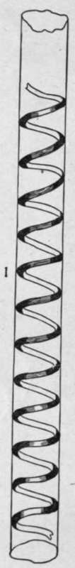
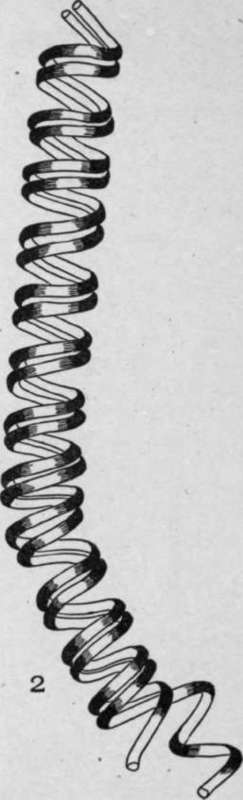
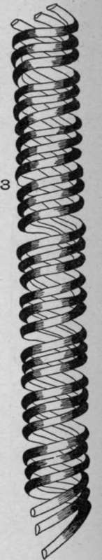
Plate 41. Spiral Vessels.
1. Single spiral vessel of pumpkin stem (Cucurbita pepo, L.).
2. Double spiral vessel of squill bulb (Urginea maritima, [L.J Baker).
3. Triple spiral vessel of eucalyptus leaf (Eucalyptus globulus, Labill).
Continue to:
My Books

