Conducting Tissue. Part 2
Description
This section is from the "Histology of Medicinal Plants" book, by William Mansfield. Also see Amazon: Histology of Medicinal Plants.
Conducting Tissue. Part 2
Sclariform Vessels
Sclariform vessels have interrupted bands of thickening on the inner walls. Two or more such bands occur between the two side walls. The series of bands are separated by uniformly thickened portions of the wall extending parallel to the length of the vessel. Sclariform vessels are usually quite broad, so that it is necessary to change the focus several times in order to bring the different series of bands in focus. The series of bands are usually of unequal width and length.
Sclariform vessels occur in male fern (Plate 42, Fig. 2), calamus, tonga root (Plate 42, Fig. 3), and sarsaparilla (Plate 42, Fig. 1). In each they are characteristic. Sclariform vessels, with these few exceptions, do not occur in drug plants. In fact, drugs derived from dicotyledones rarely have sclariform vessels. They occur chiefly in the ferns and drugs derived from mono-cotyledenous plants. Their presence or absence should, therefore, be noted when studying powdered drugs.

Plate 42. Sclariform Vessels.
1. Sarsaparilla root (Smilax officinalis, Kunth).
2. Male fern (Dryopteris marginalis, [L.] A. Gray).
3. Tonga root.
Reticulate Vessels
Reticulate vessels are of common occurrence in medicinal plants. In fact, they occur more frequently than any other type of vessel. The basic structure of reticulate vessels (Plate 43, Fig. 1) occurring in different plants is similar, but they vary in a recognizable way in different plants (Plate 43, Fig. 2). The walls of reticulate vessels are thickened to a greater extent than are the walls of spirally thickened vessels.


Plate 43. Reticulate Vessels.
1. Hydrastis rhizome (Hydrastis canadensis, L.). 2. Musk root (Ferula sumbul, [Kauffm.] Hook., f.).
Pitted Vessels
Pitted vessels are met with most frequently in woods and wood-stemmed herbs. There are two distinct types of pitted vessels - i.e., simple pitted vessels and pitted vessels with bordered pores.
The pitted vessel represents the highest type of cell-wall thickening. The entire wall of the vessel is thickened, with the exception of the places where the pits occur. The number and size of the pits vary greatly in different drugs. In quassia (Plate 44, Fig. 1) the pits are numerous and very small, and the openings are nearly circular in outline. In white sandalwood (Plate 44, Fig. 3). the pits are few in number, but when they do occur they are much larger than are the pits of quassia.


Plate 44. Pitted Vessels.
1. Quassia, low magnification (Purana excelsa, [Swartz] Lindl.).
2. Quassia, high magnification.
3. White sandalwood (Santalum album, L.).
Pitted Vessels With Bordered Pores
Pitted vessels with bordered pores are of common occurrence in the woody stems and stems of many herbaceous plants (Plate 45, Figs. 3 and 4). In such vessels the wall is unthickened for a short distance around the pits. This unthickened portion may be either circular or angled in outline, a given form being constant to the plant in which it occurs. The pits vary from oval to circular. Pitted vessels with bordered pores occur in belladonna and aconite stems.


Plate 45. Vessels.
1. Reticulate vessel of calumba root (Jateorhiza palmata, [Lam.l Miers).
2. Reticulate tracheid of hydrastis rhizome (Hydrastis canadensis, L.).
3. Pitted vessel with bordered pores of belladonna stem.
4. Pitted vessel with bordered pores of aconite stem (Aconitum napellus,L.).
Vessels and tracheids lose their living-cell contents when fully developed. In the vessels the cell contents disappear at the period of dissolution of the cell wall.
The walls of vessels and tracheids are composed of lignin, a substance which prevents the collapsing of the walls when the surrounding cells press upon them, and which also prevents the tearing apart of the wall when the vessel is filled with ascending liquids under great pressure. Lignin thus enables the vessel to resist successively compression and tearing forces.
Tracheids are formed from superimposed cells with oblique perforated end walls. The side walls of tracheids are thickened in a manner similar to those of vessels. The tracheids in golden seal are of a bright-yellow color, and groups of these short tracheids scattered throughout the field form the most characteristic part of the powdered drug. In ipecac root the tracheids are of a porcelain-white, translucent appearance, and they are much longer than are the tracheids of golden seal.
The cellulose walls of parenchyma cells are stained blue with haematoxylin and by chlorzinciodide. Cellulose is completely soluble in a fresh copper ammonia solution.
Sieve Tubes
Sieve tubes are downward-conducting cells. They conduct downward proteid food material. This fact is easily demonstrated by adding iodine to a section containing sieve tubes, in which case the sieve tubes are turned yellow.
Developing sieve tubes have all the parts common to a living cell; but when fully mature, however, the nucleus becomes disorganized, but a layer of protoplasm continues to line the cell wall.
Sieve tubes (Plate 46, Fig. 1) are composed of a great number of superimposed cells with perforated end walls and with non-porous cellulose side walls. The end walls of two adjoining cells are greatly thickened and the pores pass through both walls. This thickened part of the porous end walls of two sieve cells is called the sieve plate, and it may be placed in an oblique or a horizontal position.
In a longitudinal section the sieve tubes are seen to be slightly bulging at the sieve plate, and through the pores extend protoplasmic strands. The strands are united on the upper and lower side of the sieve plate to form the protoplasmic strands of the living sieve tubes and the callus, layers of dried plants. This callus is frequently yellowish in color, and in all cases is separated from the cell wall. In certain plants the sieve plate occurs on the side walls of the sieve tubes in contact with other sieve tubes.

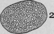
Plate 46. 1. Longitudinal section of sieve tube (Cucurbita pepo, L.).
2. Cross-section of sieve tube just above an end wall - sieve plate.
Continue to:
My Books

