Mechanical Tissues. Part 2
Description
This section is from the "Histology of Medicinal Plants" book, by William Mansfield. Also see Amazon: Histology of Medicinal Plants.
Mechanical Tissues. Part 2
Porous And Striated Bast Fibres
Porous and striated walled bast fibres occur in blackberry bark of root, wild-cherry bark, and in cinchona bark.
The fibres of blackberry root bark (Plate 23, Fig. 1) have distinctly porous and striated walls; the cavity, which is usually greater than the diameter of the wall, contains starch. These fibres usually occur as fragments.
In wild-cherry bark (Plate 23, Fig. 2) the fibre has short, thick, unequally thickened walls, which are porous and striated. Most of the fibres are unbroken.
Yellow cinchona bark (Plate 23, Fig. 3) has very thick, prominently striated porous-walled fibres, with either blunt or pointed ends. The cavity is narrow, and the pores are simple or branched.
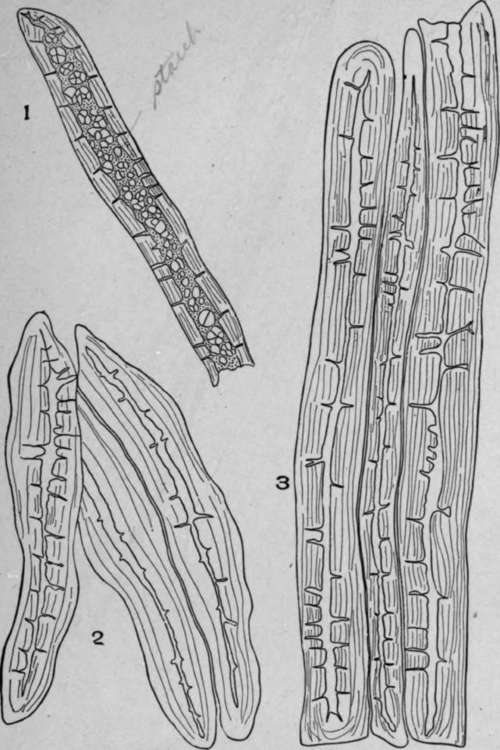
Plate 23. Porous and Striated Bast Fibres.
1. Blackberry root (Rubus cuneifolius, Pursh.).
2. Wild cherry (Prunus serotina, Ehrh.).
3. Yellow cinchona (Cinchona species).
Porous And Non-Striated Bast Fibres
Porous and non-striated bast fibres occur in marshmallow root and echinacea root.
The fibres of marshmallow (Plate 24, Fig. 3) usually occur in fragments. The walls have simple pores, and the diameter of the cell cavity is very wide; the pores on the upper or lower wall are circular or oval in outline (end view).
The bast fibres of echinacea root (Plate 24, Fig. 4) are seldom broken; the walls are yellow, the pores are simple and numerous. The edges and surface of the fibres are frequently covered with a black intercellular substance.
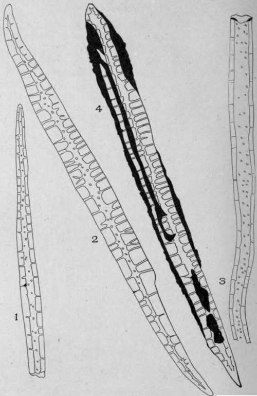
Plate 24. Porous and Non-Striated Bast Fibres.
1. Sarsaparilla root (Hypoderm), {Smilax officinalis, Kunth).
2. Unicorn root (Endoderm).
3. Marshmallow root (Althaea officinalis, L.)
4. Echinacea root (Echinacea angustifolia, D. C.).
Non-Porous And Striated Bast Fibres
Non-porous and striated bast fibres occur in elm bark, stillingia root, and cundurango bark. The bast fibres of elm bark (Plate 25, Fig. 1) occur in broken, curved, or twisted fragments. The central cavity is very small, and the walls are longitudinally striated.
In powdered stillingia root (Plate 25, Fig. 2) the bast fibres are broken, and the wall is very thick and longitudinally striated. The central cavity is small and usually not visible. Bast fibres of cundurango (Plate 25, Fig. 3) are broken in the powder. The cavity is very narrow, and the striations are arranged spirally, less frequently transversely.
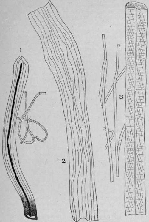
Plate 25. Non-Porous and Striated Bast Fibres.
1. Elm bark (Ulmus fulva, Michaux).
2. Stillingia root (Stillingia sylvatica, L.).
3. Cundurango root bark (Marsdenia cundurango, [Triana] Nichols).
Non-Porous And Non-Striated Bast Fibres
Non-porous and non-striated walled bast fibres occur in mezereum bark, in Ceylon cinnamon, in sassafras root bark, and in soap bark.
The simplest non-porous and non-striated walled bast fibres are found in mezereum bark (Plate 26, Fig. 4). The individual fibre is very long. If often measures over three millimeters in length, so that in the powder the fibre is usually broken. The wall is non-lignified, white, non-porous, and of uniform diameter.
In Ceylon cinnamon (Plate 26, Fig. 2) the bast fibres measure up to .900 mm. in length, so that in powdering the bark the fibre is rarely broken. These bast fibres, unlike the bast fibres of mezereum, have thick, white walls and a narrow cell cavity. Both ends of the fibre taper gradually to a long, narrow point.
In Saigon cinnamon the bast fibres are not as numerous as they are in Ceylon cinnamon. The individual fibres are thicker than in Ceylon cinnamon, and the walls are yellowish and rough and the ends bluntly pointed. These fibres are rarely ever free from adhering fragments of parenchyma tissue.
In sassafras root bark (Plate 26, Fig. 3) the fibre has one nearly straight side - the side in contact with the other bast fibres - and an outer side with a wavy outline, caused by the fibre's pressing against parenchyma cells, the point of highest elevation being the point of the fibre's growth into the intercellular space between two cells. The outer part of the wall tapers gradually at either end to a sharp point. The walls are white, thick, and non-porous.
In soap bark (Plate 26, Fig. 1) the bast fibres have thick, white, wavy walls and a narrow cavity. One end of the cell is frequently somewhat blunt while the opposite end is slightly tapering.
The branched stone cells of wild-cherry bark have three or more branches. The pores are small and usually non-branched, and the striations are very fine and difficult to see unless the iris diaphragm is nearly closed. The central cavity is very narrow and frequently contains brown tannin.
The branched stone cells of hemlock bark are very large; the walls are white and distinctly porous bordering on the cell cavity, which contains bright reddish-brown masses of tannin.
In cross-section bast fibres occur singly or isolated, as in Saigon cinnamon (Plate 34, Fig. 1); or in groups, as in meni-spermum (Plate 27, Figs. 1 and 2); or in the form of continuous bands, as in buchu stem (Plate 100, Fig. 5).
Bast fibres are seen in longitudinal view in powdered drugs. The cell cavity shows throughout the length of the fibre. This cavity differs greatly in different fibres. In soap bark (Plate 26, Fig. 1) there is scarcely any cell cavity, while in mezereum bark (Plate 26, Fig. 4) the cell cavity is very large.
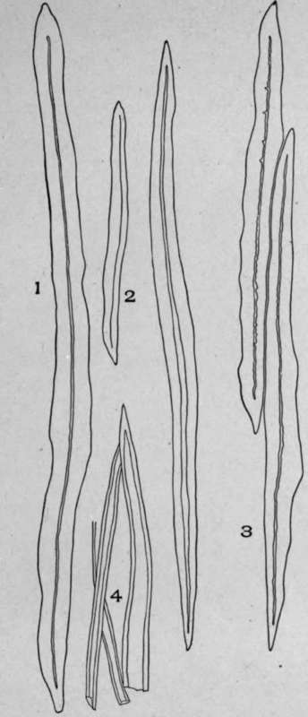
Plate 26. Non-Porous and Non-Striated Bast Fibres.
1. Soap bark (Quillaja saponaria, Molina).
2. Ceylon cinnamon bark (Cinnamomum zeylanicum, Nees).
3. Sassafras root bark (Sassafras variifolium, [Salisb.] Kuntze). $. Mezereum bark (Daphne mezereum, L.).

Plate 27. Groups of Bast Fibres.
1. Menispermum rhizome (Menispermum canadensis, L.).
2. Althea root (Althaea officinalis, L.) showing two groups of bast fibres.
The pores, which are absent in many drugs, are, when present, either simple, as in echinacea root (Plate 24, Fig. 4), or they are branched, as in yellow cinchona (Plate 23, Fig. 3).
In each of the above fibres the length and width of the fibre are shown. The fibres also have pores of variable length. Such a variation is common to most fibres with pores. That part of the wall immediately over or below the cell cavity shows the end view or diameter of the pore, as in the fibre of marsh-mallow root (Plate 24, Fig. 3). As a rule, however, the pores show indistinctly on the upper and lower wall.
Continue to:
My Books

