Mechanical Tissues. Part 3
Description
This section is from the "Histology of Medicinal Plants" book, by William Mansfield. Also see Amazon: Histology of Medicinal Plants.
Mechanical Tissues. Part 3
Occurrence In Powdered Drugs
In powdered drugs bast fibres occur singly or in groups. The individual fibres may be broken, as in mezereum and elm bark, or they may be entire, as in Ceylon cinnamon and in sassafras bark (Plate 26, Figs. 2 and 3).
The lignified walls of bast fibres are colored red by a solution of phlorogucin and hydrochloric acid, and the walls are stained yellow by aniline chloride.
In fact, few of the fibres found in individual plants occur in a broken condition.
Isolated bast fibres are circular in outline. Bast fibres, when forming part of a bundle, have angled outlines when they are completely surrounded by other bast fibres; but when they occur on the outer part of the bundle, and when in contact with parenchyma or other cortical cells, they are partly angled and partly undulated in outline.
In the bast fibres the pores are placed at right angles to the length of the fibre. The side walls show the length of the pore (Plate 24, Fig. 3); while the upper or lower wall shows the outline, which is circular, and the pore, which is very minute.
Most bast fibres have no cell contents. In some cases, however, starch occurs, as in the bast fibres of rubus.
The color of the bast fibres varies, being colorless, as in Ceylon cinnamon; or yellowish-white, as in echinacea; or bright yellow, as in bayberry bark.
Bast fibres retain their living-cell contents until fully developed; then they die and function largely in a mechanical way.
The walls of bast fibres are composed of cellulose or of lignin. Most of the bast fibres occurring in the medicinal plants give a strong lignin reaction.
Wood Fibres
Wood fibres always occur in cross-sections associated with vessels and wood parenchyma, from which they are distinguished by their thicker walls, smaller diameter, and by the nature of the pores, which are usually oblique and fewer in number than the pores in the walls of wood parenchyma, and different in form from the pores of vessels.
The wood fibre on cross-section (Plate 105, Fig. 4) shows an angled outline, except in the case of the fibres bordering the pith-parenchyma, etc., in which case they are rounded on their outer surface, but angled at the points in contact with other fibres. The pore of wood fibres is one of the main characteristics which enable one to distinguish the wood fibres from bast fibres.
The pores are slanting or strongly oblique (Plate 28, Fig. 2), and they show for their entire length on the broadest part of the wall - i.e., the upper or the lower surface - while in the side wall they are oblique; but they are not so distinct as they are on the broad part of the wall.
Frequently the pores appear crossed when the upper and the lower wall are in focus, because the pores are spirally arranged, and the pore on the under wall throws a shadow across the pore on the upper wall, or vice versa.
Wood fibres always occur in a broken condition (Plate 28, Fig. 1) in powdered drugs. These broken fibres usually occur both singly and in groups in a given powder.
The color of wood fibres varies greatly in the different medicinal woods. Fragments of wood are usually adhering to witch-hazel, black haw, and other medicinal barks. In each of these cases the wood fibres are nearly colorless. In barberry bark adhering fragments of wood and the individual fibres are greenish-yellow. The wood fibres of santalum album are whitish-brown; of quassia, whitish-yellow; of logwood and santalum rubum, red.
Some wood fibres function as storage cells. In quassia the wood fibres frequently contain storage starch. The wood fibres of logwood and red saunders contain coloring substances, which are partially in the cell cavity and partially in the cell wall. The walls of wood are composed largely of lignin.

Plate 28. Wood Fibres.
1. White sandalwood (Santalum album, L.).
2. Quassia wood {Picraena excelsa, [Swartz] Lindl.).
3. Logwood with crystals (Hamatoxylon campechianum, L.)
4. Black haw root (Viburnum prunifolium, L.).
Collenchyma Cells
Collenchyma cells form the principal medicinal tissue of stems of herbs, petioles of leaves, etc. In certain herbs the collenchyma forms several of the outer layers of the cortex of the stem. In motherwort, horehound, and in catnip the collenchyma cells occur chiefly at the angles of the stem. In motherwort (Plate 29, Fig. B) there are twelve bundles, one large bundle at each of the four angles, and two small bundles, one on either side of the large bundle. In catnip (Plate 29, Fig. A) there are four large masses, one at each angle of the stem.
Collenchyma cells differ from parenchyma cells in a number of ways: first, the cell cavity is smaller; secondly, the walls are thicker, the greater amount of thickening being at the angles of the cells - that is, the part of the cell wall which is opposite the usual intercellular space of parenchyma cells, while the wall common to two adjoining cells usually remains unthickened. In horehound stem (Plate 30, Fig. 2) the thickening is so great at the angles that no intercellular space remains. In the side column of motherwort stem (Plate 30, Fig. 1) the thickening between the cells has taken place to such an extent that the cell cavities become greatly separated and arranged in parallel concentric rows.
The collenchyma of the outer angle of motherwort stem (Plate 30, Fig. 3) is greatly thickened at the angles. There are no intercellular spaces between the cells, and cell cavity is usually angled in outline instead of circular, as in the cells of horehound. In certain plants intercellular spaces occur between the cells, and the walls are striated instead of being non-striated, as in the stems of horehound, motherwort, and catnip.
Collenchyma cells retain their living contents at maturity. Many collenchyma cells, particularly of the outer layers of bark and the collenchyma of the stems of herbs, contain chlorophyll.

Plate 29. A. Diagrammatic sketch of the cross-section of catnip stem (Nepeta cateria, L.). I. Collenchyma occurring at the four angles of the stem.
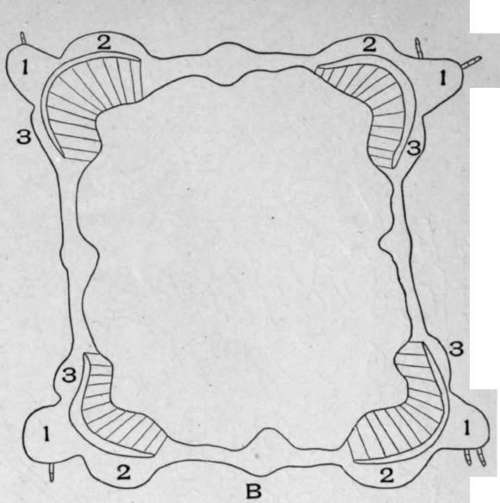
B. Diagrammatic sketch of the cross-section of motherwort stem (Leonurus cardiaca, L.). 1, 2, 3. Twelve masses of collenchyma tissue occurring at the four sides of the stem.

Plate 30. Collenchyma Cells.
1. Cross-section of a side column of the collenchyma of motherwort stem (Leonurus cardiaca, L.).
2. Cross-section of the collenchyma of horehound stem (Marrubium vulgare, L.).
3. Cross-section of the collenchyma of the outer angle of motherwort stem.
The walls of collenchyma consist of cellulose.
Continue to:
My Books

