


Natural Mating, Equipment For Instrumental Insemination, Microscope And Light, Carbon Dioxide Equipment, Queen Holder
Description
This section is from the book "Instrumental Insemination Of Queen Bees", by Otto Mackensen, Kenneth W. Tucker. Also available from Amazon: Instrumental Insemination Of Queen Bees.
Natural Mating, Equipment For Instrumental Insemination, Microscope And Light, Carbon Dioxide Equipment, Queen Holder
Natural Mating
The natural act of copulation has been difficult to study, but considerable information has been obtained by examining queens immediately after their return from the mating flight. The queen evidently cooperates actively by opening the sting chamber and lowering the valvefold voluntarily. The end of the penis enters the bursa copulatrix but not the vagina. Since the queen is not cooperating during artificial insemination, it may be necessary to lower the valvefold with a probe, to facilitate placement of the syringe tip in the median oviduct.
When the queen returns to the hive after mating, usually both oviducts are distended with semen, but often unequally. The anterior part of the vagina also contains semen, and some sperm have already reached the spermatheca. The bulb of the penis is found in the sting chamber or bursa copulatrix buried in mucus which also extends into the vagina, or the bulb may be absent.
During the course of the next 6 to 7 hours most of the sperm reach the spermatheca through the vagina and spermathecal duct, but some may remain in the oviducts 24 hours, especially after artificial insemination. How this translocation takes place is not yet perfectly understood. It has long been thought to be a process of active migration; however, while it is taking place, the queen frequently contracts her abdomen and little strings of dried semen are eliminated. From this evidence it has been assumed that the sperm is forced into the spermatheca during these contractions while the valvefold serves as an imperfect seal of the vaginal orifice. Perhaps both processes play a part. Whatever the procedure, it is as effective after instrumental insemination as after natural mating.
Naturally mated queens, on return from mating flights, contain an average of 11. 6 microliters (microliter) of semen in their oviducts (maximum 28 microliter ). From this it is estimated that queens mate with eight to nine drones on the average (maximum: 17). If the first mating does not satisfy, the queen will mate a second or even a third time.
Equipment For Instrumental Insemination
The major equipment needed to perform the insemination operation is illustrated in figure 5. It consists of the manipulating apparatus (fig. 6) to which the queen holder (QH), ventral hook (VH), and sting hook (STH) and syringe (S) are attached in movable fashion, a cylinder of carbon dioxide for use as an anesthetic, a stereomicroscope, and a source of light. A jar in which queens are given additional treatments with carbon dioxide is also shown.
Microscope And Light
The microscope should be of the stereoscopic type with a wide field. The most desirable magnifications will vary with the operator. An experienced operator may prefer to use a single magnification of 10 to 15 diameters for both the collection of semen and insertion of the syringe. A beginner may wish to have also a higher magnification of about 20 diameters available for the insertion; the experienced operator might find 20 diameters useful with difficult queens. The experienced operator may be able to use an even lower magnification (6 to 7 diameters) so that the two operations can be performed under a single field without moving the microscope.
Some type of illumination is essential. A microscope lamp with a concentrated beam of adjustable brightness is best, but any lamp that gives sufficient light to satisfy the operator is satisfactory. The beam should be large enough to cover both the semen collection and injection operations without moving the lamp, or the lamp should be attached to the microscope so the point of focus will always remain within the beam.
Carbon Dioxide Equipment
Carbon dioxide serves as anesthetic. It is obtainable in cylinders under high pressure which must be reduced to a few pounds per square inch by a reduction valve. If taken directly from the cylinder the gas will alternately freeze and thaw causing an irregular flow of gas. A needle valve permits adjustment of the flow to a very fine stream. A rubber tube carries the gas to the queen holder and another tube leads to a container in which queens are given additional anesthetizations.
Queen Holder
The queen holder (fig. 7) consists of an outer transparent plastic tube 1 1/2 inches long and an inner stopper (also a tube) of the same length to which the carbon dioxide supply tube is attached. The stopper is made of two parts that screw together permitting the insertion of a felt washer. This washer provides friction to keep the stopper in place and prevents leakage of carbon dioxide back around the stopper. The friction is adjustable to a limited extent. Screwing the parts together more tightly tends to increase the diameter of the felt washer thus increasing the friction with the tube. The queen is induced to back into this tube and when her abdomen begins to protrude she is secured with the stopper. The carbon dioxide flows through the hole in the stopper and bathes the queen as it passes out of the queen holder.
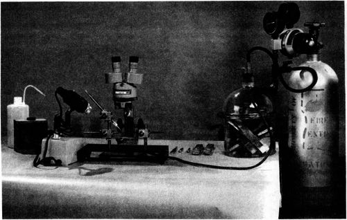
Figure 5. -Complete insemination equipment, showing manipulating apparatus under microscope with queen in place ready for injection of semen, jar for giving additional carbon dioxide treatments, and carbon dioxide cylinder with pressure regulator attached.
Continue to:


My Books

