Diseases Of The Mammary Gland
Description
This section is from the book "A Manual Of Pathology", by Guthrie McConnell. Also available from Amazon: A Manual Of Pathology.
Diseases Of The Mammary Gland
Malformations
As an associated condition with imperfect development of the chest-walls one or both glands may be absent. They may be hypoplastic, when there is also an incomplete development of the sexual organs. A breast may be normal in other respects, but be lacking a nipple, or there may be several nipples. Supernumerary mammae - polymastia - may be present in both sexes, usually on the anterior surface of the chest and abdomen. They may occur on the back or thigh, and occasionally may functionate, although they are usually ill developed and lack a nipple.
Circulatory Disturbances
Hyperemia is present during menstruation, during pregnancy, and at the beginning of lactation. The gland will be reddened, swollen, and sometimes painful. This congestion may also be brought about by some diseased condition of the uterus, the relationship of these two organs being very close.
Hemorrhage is due to some injury of the gland. The bleeding may take place within the connective tissue, into the glandular structures, or deeper down, behind the gland upon the muscle. The blood may escape from the nipple, it may be absorbed, or it may become encapsulated by a wall of fibrous tissue and form a hematoma. Hemorrhage may also be the result of bleeding from the ulcerated surfaces of new growths.
Inflammation of the mammae, or mastitis, may rarely be due to injury, but it is most commonly the result of infection occuring during the puerperium. The micro-organisms most frequently gain entrance through fissures of the nipple during suckling. Infection directly into the milk-ducts is not common. Mastitis may result from the extension of inflammations of neighboring structures, as caries of a rib, erysipelas of the skin, or in puerperal infection the micro-organisms may have been brought to the gland through the blood-vessels. The disease may be diffuse or involve a portion only of the gland, the latter being the more common. In the diffuse form the inflammation may extend to neighboring structures, setting up a paramastitis. In the circumscribed variety there is abscess formation, which may be single or multiple. The pus may escape into the milk-ducts and out through the nipple; it may be interstitial or rupture externally, in the latter case frequently causing a fistula. If the pus burrow into the deeper tissues, it may perforate into the pleural cavity and cause a fatal empyema. Occasionally the pus may be encapsulated, inspissated, and calcified. Sometimes a condition of the mammae similar to that of chronic interstitial inflammation of the organs occurs. The glands are firm and hard, due to the connective-tissue formation, and small cystic dilatations of obstructed milk-ducts may be present.
Tuberculosis of the mammary glands is rare, except as a secondary involvement in tuberculosis of the axillary nodes or other tissues. The tubercle bacilli are probably carried by the blood. Tubercles are formed which undergo caseation, and the contents escape into the acini. In this way great numbers of the bacilli can gain entrance into the milk.
Syphilis of the mammae is very rare, but has been seen as gummata, which in healing form a dense, stellate scar.
Atrophy of the glands occurs after the menopause or when the ovaries have been removed.
Hypertrophy at the time of puberty may continue beyond the normal limits and cause an enormous development of both the glandular and connective-tissue elements of either or both breasts. If lactation takes place, the amount of milk secreted may be very great. The gland may be much enlarged, on account of a diffuse fatty infiltration or lipomatosis.
Tumors
Sarcoma is rather infrequent, is usually of the round-cell variety, and may be diffuse or in circumscribed nodules. One gland alone is generally involved. In the diffuse form the mamma rapidly enlarges, the growth infiltrates in all directions, the skin soon becomes firmly attached and may ulcerate. The structure of the tumor differs in different parts. It may be quite cystic on account of obstruction to the milk-ducts; part may be myxomatous or resemble connective-tissue. The sarcoma cells may extend into the cystic dilatations as polypoid projections - intracanalicular sarcoma.
Occasionally the tumors may be circumscribed. They, most commonly originate within the adventitia of the milk-ducts and nipple, but may arise from any part of the connective-tissue of the gland. These tumors give metastasis by means of the blood, but they are much less malignant than the carcinomata.
Fibroma as a pure connective-tissue tumor is unusual. It is commonly found in connection with a hyperplasia of the glandular structures.
Adenoma in a typical form is rare, is generally associated with an overgrowth of connective-tissue, and is called either adenofibroma or fibro-adenoma. According to the relation of the glandular and fibrous elements these tumors can be classified into three divisions:
Inter canalicular fibro-adenoma, in which the tumor is chiefly fibrous in structure, with the ducts and acini irregularly distributed through it.
"Pericanalicular fibro-adenoma, in which the fibrous tissue makes distinct concentric investments of the ducts and groups of acini.
" Intracanalicular fibro-adenoma, in which polypoid or papillary growths extend into the ducts".
These tumors are more or less completely encapsulated, and although of the benign type, they not infrequently take on a carcinomatous growth.
Carcinoma is an extremely common tumor of the mammary gland in women between the ages of forty and fifty. About 2 per cent, of cases occur in males. The growth usually involves one breast only, and that the right more frequently than the left. It develops either from the tubules or the acini of the glands, and may start as a carcinoma or result from a malignant degeneration of a fibro-adenoma. When the growth begins in the acini, it resembles quite closely the ordinary racemose character of the gland - is alveolar in form. If it is of the tubular type, there are long tubular collections of cells.
Although at first the growth may quite closely resemble a simple adenoma, proliferative changes soon occur in the epi-theuum. The cells, instead of forming a single layer, increase in number and lose their resemblance to the normal structure. These new-formed cells may completely fill the acini, or they may be found within the surrounding tissue as a result of destruction of the basement membrane.

Fig. 178.
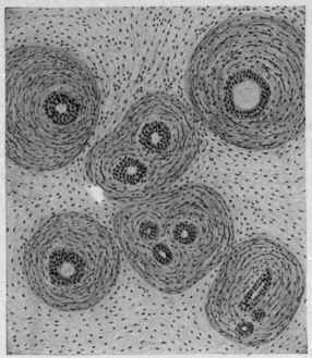
Fig. 179.
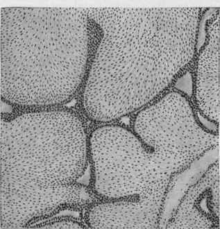
Fig. 178. - Intercanalicular adeno-fibroma of mamma. The fibro-con-nective tissue bears no definite relation to the glandular canals. Fig. 179. - Pericanalicular adeno-fibroma of mamma. The fibro-connective tissue shows a peculiar concentric relation to the glandular canals. Fig. 180. - Intracanalicular adeno-fibroma of mamma. Papillary connective-tissue growths project into the glandular canals (McFarland).
Fig. 180.
According to the relationship between stroma and parenchyma mammary carcinoma may be divided into three classes:
Carcinoma simplex, in which there is, relatively speaking, an equal amount of connective-tissue and epithelial cells. Is less malignant than the medullary, but more so than the scirrhous.
Encephaloid or medullary carcinoma, which is very rich in cells and poor in stroma, is soft, contains much "cancer juice," and hemorrhages are not uncommon. It grows rapidly, soon ulcerates, and is rapidly fatal.
Scirrhous carcinoma is characterized by a great preponderance of connective tissue, is hard, is slow in growth, does not tend to give metastases, and is slowly fatal. In this form there is frequently a retraction of the nipple.
The so-called colloid carcinoma of the breast is generally one in which a myxomatous degeneration of the connective tissue has given rise to the appearance.
Extension of carcinoma may take place directly to the skin or, by penetrating deeply, enter the chest-walls and pleurae. Metastasis is common, takes place early, and may be very extensive, the axillary nodes being first involved. Secondary growths may also occur in distant parts of the body.
Paget's disease begins as a chronic eczema of the nipple and adjacent skin. It may have existed for ten or fifteen years with more or less complete destruction of the nipple, and then take on a carcinomatous change.
Cysts are quite commonly found, particularly in new growths in which there has been an attempt at secretion without any outlet. Small milk-cysts may result from obstruction to the ducts. The contents of the cysts may be milky, or, through absorption of the liquid portion, become thick and caseous. In some cases there are polypoid outgrowths of connective tissue and epithelium from the walls of the cysts.
Continue to:
My Books

