Infectious Diseases Of The Liver
Description
This section is from the book "A Manual Of Pathology", by Guthrie McConnell. Also available from Amazon: A Manual Of Pathology.
Infectious Diseases Of The Liver
Tuberculosis of the liver is secondary to lesions of the disease elsewhere, particularly of the peritoneum, and is probably transmitted through the blood. It appears generally as miliary tubercles scattered throughout the organ, or as larger necrotic foci. Rarely there is a single large cheesy focus.
Microscopically the lesions are the same typical ones as are found everywhere in the disease. Such a liver is usually fatty, but may show amyloid changes.
Syphilis of the liver is a common involvement in that disease. In adults who have acquired syphilis there is frequently a diffuse proliferation of connective tissue with atrophy of the hepatic cells that closely resembles atrophic cirrhosis. Generally the disease manifests itself in the form of localized proliferations of connective tissue that divide the liver into numerous small but well-defined lobes. This results from the T. pallidum getting into the circulation, becoming lodged in the minute vessels and producing a toxin that causes the hyperplasia. A certain area may become almost constricted off from the rest of the organs. The irregular distribution of the connective tissue is the characteristic feature. This form probably originates as an inflammatory thickening about the portal veins and the bile-ducts.
Gumma may also be present in acquired or congenital syphilis, either singly or in numbers. In the acquired form the single ones are usually the larger. They occur as rounded yellowish masses, the center of the larger ones frequently being the seat of coagulation necrosis. Surrounding them is a zone of hyperemia and there is generally some connective-tissue hyperplasia. This occurs in the form of bands radiating from the center, giving a characteristic stellate appearance to the resulting scar.
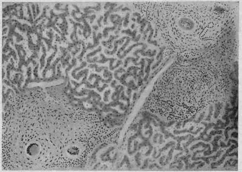
Fig. 160. - Miliary Tuberculosis of the Liver. X 70 (Dürck).
Two foci, consisting of smaller confluent tubercles, which are still distinguishable. The giant cells are rounded. The foci are situated in the periportal tissue in the vicinity of a portal branch.
Congenital syphilis of the liver may manifest itself in a diffuse form or as gummata. In the diffuse variety there is a wide-spread connective-tissue proliferation, resembling that of biliary cirrhosis, and round-cell infiltration. The organ is yellowish or brown, sometimes larger than normal, and extremely firm, almost like sole leather. The round-cell infiltration is found in the neighborhood of the bloodvessels, even being seen within the walls. The liver epithelium is frequently the seat of fatty degeneration, giving rise to the "acute yellow atrophy" of the liver of the newborn.
The congenital gummata are not, as a rule, circumscribed. They are found in the interlobular tissue and may be located within the wall of a blood-vessel or of a bile-duct.
Leprosy is sometimes found in the liver, where it occurs as granulomatous masses containing the characteristic giant cells and bacilli.
Actinomycosis rarely occurs in the liver. When present, it is generally due to secondary involvement by extension from the lung.
Continue to:
My Books

