Physical Examination of Breasts
Description
This section is from the book "Early Detection And Diagnosis Of Cancer", by Walter E. O'Donnell. Also available from Amazon: Early Detection And Diagnosis Of Cancer.
Physical Examination of Breasts
Good light, preferably from a source located behind, above, and to one side of the examiner, is essential. The patient is uncovered to the waist. 1. With the patient sitting
(a) Inspect the breasts for evidence of masses, asymmetry, or nipple abnormality (e.g., ulceration, inversion, discharge)
(1) With the patient's arms at her sides (Fig. 12, A)
(2) With the patient's arms extended overhead (Fig. 12, B)
(b) Palpate for evidence of adenopathy in the
(1) Axillae, with the patient's elbow cupped in your hand (Fig. 13)
(2) Supraclavicular areas (Fig. 14)

Fig. 12. Inspection of the breasts with the patient sitting. A, With the arms at the sides. B, With the arms extended overhead.
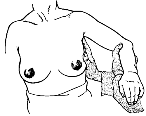
Fig. 13. Palpation of the axilla with the patient sitting.
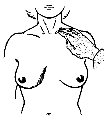
Fig. 14. Palpation of the supraclavicular areas with the patient sitting.

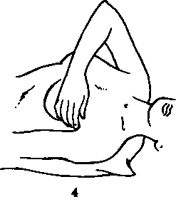
Fig. 15. Palpation of the breasts with the patient lying down. A, With the arm at the side. B, With the arm behind the head.
2. With the patient lying down
(a) Palpate the breasts
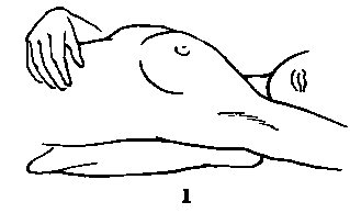
(1) With the patient's arm at her side (Fig. 15, A)
(2) With the patient's arm behind her head (Fig. 15, B )
A small pillow may be placed under the chest on the side being examined to promote relaxation of the breast and supporting structures.
(b) Technique
(1) Palpation is best accomplished by the flat surface or balls of the fingers of both hands. A gentle touch is more sensitive and informative than needless massaging and kneading of breast tissue between the fingers.
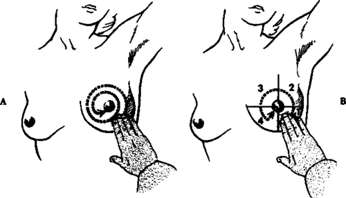
Fig. 16. Technique of breast examination. A, Starting at the periphery and working toward the nipple in ever-decreasing concentric circles. B, Examining each quadrant separately in sequence.
(2) The entire surface of each breast, especially the vulnerable outer quadrants and tails, must be covered.
(3) Some start out in the periphery of the breast and work toward the nipple in ever-decreasing concentric circles (Fig. 16, A)
(4) Others prefer to think of the breast in terms of quadrants and examine each of these four sectors completely in turn (Fig. 16, B)
(5) The nipple and subareolar tissue must be carefully palpated; a gentle attempt may be made to express secretion from the nipple, especially if there is a history of previous discharge or bleeding.
(6) The undersurface of the breast must be given special attention in the recumbent position, since when the patient is sitting up it is hidden from view; furthermore, normal structures constituting the inframammary ridge may be mistaken for significant lesions.
(7) The exact details of technique are less important than the fact that the examination is complete, painstaking, and systematic.
3. Instruct the patient in breast self-examination
The rationale, as well as the pros and cons, of indoctrination in the habit of breast self-examination is discussed in the separate chapter on breast cancer. Only the technique will be noted here.
(a) With the patient sitting
Demonstrate to the patient how she should sit or stand in a good light, facing a mirror, uncovered to the waist, and inspect her breasts -first with the arms relaxed at her side and then with the arms overhead (Fig. 17).

Fig. 17. Breast self-examination. The patient inspects her breasts in a mirror with arms at side, A, and with arms elevated, B.

Fig. 18. Instruction in breast self-examination with the patient lying down. In this illustration the patient is guided in palpating her right breast with her left hand. The procedure is then repeated on the left breast with the right hand.
(b) With the patient lying down
Guide the patient's hand as you show her how to examine her breasts (Fig. 18).
The technique of breast self-examination is shown in Fig. 19. Leaflets illustrating and describing the technique are available from the various medical societies and local and state health departments for distribution to patients. However, the leaflets should be given to patients after they have been indoctrinated by a physician. In selected instances a well-trained nurse can instruct patients in the technique of breast self-examination.
The foregoing is concerned with the detection of breast cancer in females. It should be emphasized that the male breast should always be palpated also.
Lie down on a bed. Place a small flat pillow or a folded bath towel under the loft shoulder. Raise the left arm over the head and rest it on the bed.

Place the flat of the flngers of the right hand at tho breast bone. Using Arm gentle pressure, feel all parts of the upper inner quarter of the left breast, progress In toward tho nlpple, and include the area beneath the nipple.

Next, In the same manner, examine the lower Inner quarter of the left breast. Note the ridge of firm tissue or flesh. This is normal.
Place the left arm down at the side. Still using the flat of the flngers, feel the tissues up into the left armpit.


Examine the upper outer quarter of the left Examine the lower outer quarter of the left breast.
Shift tho pillow or towel to the right side and repeat tho procedure on tho right broast.
Fig. 19. Technique of breast self-examination. (Adapted from A Monthly Check, New York State Department of Health.).
Continue to:
My Books

