Physical Examination of Female Genitalia
Description
This section is from the book "Early Detection And Diagnosis Of Cancer", by Walter E. O'Donnell. Also available from Amazon: Early Detection And Diagnosis Of Cancer.
Physical Examination of Female Genitalia
With the patient in the customary lithotomy position and with the headlight used as a source of illumination, the examination is carried out as follows:
1. Inspect the external genitalia, urethral meatus, etc.
2. Obtain the vaginal smear.
(a) Insert the angled end of the glass aspirator or pipette into the posterior fornix with the rubber bulb compressed (Fig. 21, A).
(b) Release pressure on the bulb while moving the tip from side to side in the fornix, thus aspirating the pooled secretions from the vagina.
(c) Remove the pipette from the vagina.
(d) Compress the bulb and express the secretion onto a clean glass slide (Fig. 21, B, top).
(e) Spread the secretion thinly and evenly over the slide with the side of the pipette (Fig. 21, B, bottom).
(f) Immediately plunge the slide into a bottle containing a fixative (equal parts of ether and 95% alcohol). An ordinary paper clip attached to the slide will prevent it from adhering to the cervical smear slide (Fig. 21, C).
(g) A cotton-tipped applicator may be used to collect the vaginal specimen if a pipette is not available or suitable.
3. Examine the cervix and vagina with a speculum.
(a) Do not use any lubricating jelly on the speculum.
(b) If the vaginal mucosa is dry or atrophic, run warm water over the speculum and shake off the residue. This permits easier insertion of the instrument.
(c) Insert the speculum into the vagina and visualize the cervix.
(d) Observe the surface of the cervix for any abnormalities.
(e) The vaginal mucosa can be visualized in its entirety by rotation of the speculum.
4. Obtain the cervical smear.
(a) Using an ordinary cotton-tipped applicator, obtain a cervical smear by swabbing the entire surface of the cervix. Most important is twirling or rotating the applicator in the cervical os (Fig. 22, A). Spread the secretion on a clean glass slide (Fig. 22, B) and immerse the slide in the specimen bottle with the vaginal smear (Fig. 22, C). Specially designed spatulas, etc. are available for taking the cervical smear. These are not essential for the procurement of satisfactory specimens.

Compress the bulb. Introduce the aspirator along tho posterior vaginal wall. Release the bulb while moving tho tip from side to side in tho fornix.
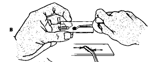
Express aspirated material on a clipped slide. Spread quickly with the side of the pipette.

Put the slide into a fixative Immediately. After one hour, slides may be removed for staining or mailing.
Fig. 21. Technique used in obtaining a vaginal smear.

Introduce the speculum without a lubricant. Inspect the vagina and cervix. Swab the entire surface of the cervix and then rotate tho swab in tho external
OS.

Spread tho material by firmly rolling the swab along the slide.

Immediately immerse the slide in the specimen bottle which contains the vaginal smear. After ana hour the slide may bo removed for staining or mailing.
Fig. 22. Technique used in obtaining a cervical smear.
(b) If much tenacious mucus is present, especially in the cervical os, some of this may be removed with a cotton swab before the smear is taken.
(c) A cervical smear should always be taken no matter how normal the cervix appears.
(d) The vaginal and cervical smears, together with complete clinical information for the cytologist, should then be sent to the laboratory.

Fig. 23. Paint the cervix with iodine-the modified Schiller test.

Fig. 24. Bimanual pelvic examination.
5. Do a modified Schiller test.
(a) Using a cotton-tipped applicator, paint the cervix with 1% tincture of iodine* (Fig. 23).
(b) The entire cervix should stain a uniform mahogany brown in premenopausal patients. In postmenopausal women the iodine stain is normally much lighter.
*Strong Iodine Tincture, N.F., prepared as follows: iodine, 7 Cm.; potassium iodide, 5 Cm.; purified water, 5 ml.; 95* ethyl alcohol qs ad 100 ml.
(c) On extremely rare occasions the iodine test is contraindicated because of iodine sensitivity, particularly in naturally blond and red-haired women, who should be queried in regard to this possibility beforehand.
(d) Determination of protein-bound iodine levels will be valueless for months after the iodine staining test is performed.
(e) Note areas which stain poorly or not at all as sites of probable future biopsy.
6. Remove the speculum.
7. Do the regular bimanual pelvic examination, noting the size, position, contour, mobility, and consistency of the uterus and palpating the cervix (Fig. 24). The adnexal regions should also be palpated and particular attention given to the possibility of any ovarian abnormalities.
8. Do a rectovaginal examination, especially when the uterus is retroverted.
Continue to:
My Books

