X-Ray Studies of Of The Colon And/Or Rectum Cancer
Description
This section is from the book "Early Detection And Diagnosis Of Cancer", by Walter E. O'Donnell. Also available from Amazon: Early Detection And Diagnosis Of Cancer.
X-Ray Studies of Of The Colon And/Or Rectum Cancer
A few additional statements are in order on the subject of x-ray studies in tumors of the rectum and colon.
1. The conventional barium enema is not sufficient by itself for the detection or diagnosis of polypoid lesions, especially the small ones. It must be supplemented and complemented by air contrast studies.
According to this technique, after barium is introduced under fluoroscopic guidance and regular films are taken, it is evacuated and the bowel insufflated with air. The dark, air-containing lumen then stands in stark contrast to the whitish, barium-coated mucosa. In an ideal study any polyps will stand out in sharp relief as whitish protuberances with a pedicle.
The air contrast study should be specifically requested by the physician when he wants to rule out polyps.
2. A gastrointestinal series or barium swallow can usually contribute little or nothing to the evaluation of disease of the rectum and colon. In the presence of an even partially obstructing lesion of the bowel, the orally ingested barium can become inpacted and cause a major problem in elimination; occasionally surgery is required. If both studies are required, the barium enema should precede the gastrointestinal series.
*These arguments deal with the question of whether or not biopsy should be accomplished by the first physician to see the patient. Total or partial biop.y and pathologic examination must ultimately be done by somebody in all cases.
A

B

C

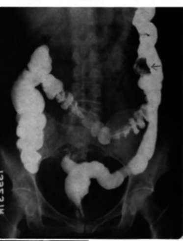
Fig. 72. A, Double contrast barium enema study showing a polyp measuring 1.5 cm. in the sigmoid colon. It may be clearly seen as a positive shadow.
B, On this upright double contrast barium enema study, a polyp on a stalk may be seen in the midsigmoid colon.
C, An unusual cluster of polypoid tumors in the midascending colon may be seen clearly outlined by positive and negative shadows in the double contrast study. Note the many diverticuli in the descending colon and note that none of the polypoid lesions protrudes outside the lumen of the bowel.
D

E
F

Fig. 72-cont'd. D, In this barium enema study it would appear that the right colon is negative. Actually, however, neither the cecum nor the terminal ileum is filled by the retrograde flow of barium because of an obstructing carcinoma of the cecum.
E, A 4 cm. ovoid filling defect in the distal portion of the transverse colon due to a polypoid tumor.
F, A 1 cm. ovoid positive shadow on the medial wall of the iliac portion of the descending colon outlined by a double contrast barium enema.
The physician may find himself confronted with a need for further work-up in one of several typical situations:
1. A lesion may be detected on routine sigmoidoscopy in an asymptomatic individual.
2. On x-ray examination a high-lying polyp may be found in the colon above the reach of the sigmoidoscope. The patient may or may not be symptomatic.
3. A patient may be suspected of having cancer of the rectum or colon as a result of symptoms.
These situations will be considered individually.
If a lesion is detected in an asymptomatic individual on routine sigmoidoscopy:
1. Describe the lesion precisely according to its:
(a) Size in millimeters
(b) Distance from anal margin in centimeters
(c) Location
(1) By relation to the face of a clock

(2) By quadrants
(d) Appearance
(1) Sessile or pedunculated
(2) Vascularity
(3) Color
(4) Etc.
2. Search carefully for other lesions. They are frequently multiple.
3. Do a barium enema x-ray study with air contrast technique in all cases for the detection of a possible high-lying lesion beyond the reach of the sigmoidoscope.
About 6% of polyps detected by sigmoidoscopy are associated with lesions in the right or left colon accessible only to x-ray technique. If a high-lying lesion is found, handle in the manner detailed below.
4. Biopsy. As noted previously, this can be done by the practitioner or consultant, according to the particular circumstances.
(a) Ideally, all polyps should be removed in toto initially in order to provide the pathologist with complete material for microscopic study. Partial biopsy of larger lesions, however, may sometimes be advisable in order to determine the nature and extent of surgery required. In general, lesions 8 to 10 mm. in size or less should be removed completely.
(b) Lesions that appear quite vascular or that are situated high in the rectum or in the rectosigmoid are often best removed in the hospital, with the patient kept at least overnight for observation.
Continue to:
My Books

