How To Use The Microscope. Part 2
Description
This section is from the "Histology of Medicinal Plants" book, by William Mansfield. Also see Amazon: Histology of Medicinal Plants.
How To Use The Microscope. Part 2
Care Of The Microscope
If possible, the microscope should be stored in a room of the same temperature as that in which it is to be used. In any case, avoid storing in a room that is cooler than the place of use, because when it is brought into a warmer room, moisture will condense on the ocular objectives and mirrors.
Before beginning work remove all moisture, dust, etc., from the inner and outer lenses of the ocular, the objectives, the Abbe condenser, and the mirror by means of a piece of soft, old linen. When the work is finished the optical parts should be thoroughly cleaned.
If reagents have been used, be sure that none has got on the objectives or the Abbe condenser. If any reagent has got on these parts, wash it off with water, and then dry them thoroughly with soft linen.
The inner lenses of the eye-pieces and the under lens of the Abbe condenser should occasionally be cleaned. The mechanical parts of the stand should be cleaned if dust accumulates, and the movable surfaces should be oiled occasionally. Never attempt to make new combinations of the ocular or objective lenses, or transfer the objectives or ocular from one microscope to another, because the lenses of any given microscope form a perfect lens system, and this would not be the case if they were transferred. Keep clean cloths in a dust-proof box. Under no circumstances touch any of the optical parts with your fingers.
Preparation Of Specimens For Cutting
Most drug plants are supplied to pharmacists in a dried condition. It is necessary, therefore, to boil the drug in water, the time varying from a few minutes, in the case of thin leaves and herbs, up to a half hour if the drug is a thick root or woody stem. If a green (undried) drug is under examination, this first step is not necessary.
If the specimen to be cut is a leaf, a flower-petal, or other thin, flexible part of a plant, it may be placed between pieces of elder pith or slices of carrot or potato before cutting.
Short Paraffin Process
In most cases, however, more perfect sections will be obtained if the specimens are embedded in paraffin, by the quick paraffin process, which is easily carried out.
After boiling the specimen in water, remove the excess of moisture from the outer surface with filter paper or wait until the water has evaporated. Next make a mould of stiff cardboard and pour melted paraffin (melting at 50 or 60 degrees) into the mould to a height of about one-half inch, when the paraffin has solidified. This may be hastened by floating it on cool or iced water instead of allowing it to cool at room temperature.
The specimens to be cut are now placed on the paraffin, with glue, if necessary, to hold them in position, and melted paraffin poured over the specimens until they are covered to a depth of about one-fourth of an inch. Cool on iced water, trim off the outer paraffin to the desired depth, and the specimen will be in a condition suitable for cutting.
Good workable sections may be cut from specimens embedded by this quick paraffin method. After a little practice the entire process can be carried out in less than an hour. This method of preparing specimens for cutting will meet every need of the pharmacognosist.
Long Paraffin Process
In order to bring out the structure of the protoplast (living part of the cell), it will be necessary to begin with the living part of the plant and to use the long paraffin method or the collodion method.
Small fragments of a leaf, stem, or root-tip are placed in chromic-acid solution, acetic alcohol, picric acid, chromacetic acid, alcohol, etc., depending upon the nature of the specimen under observation. The object of placing the living specimen in such solutions is to kill the protoplast suddenly so that the parts of the cell will bear the same relationship to each other that they did in the living plant, and to fix the parts so killed. After the fixing process is complete, the specimen is freed of the fixing agent by washing in water. From the water-bath the specimens are transferred successively to 10, 20, 40, 60, 70, 80, 90, and finally 100 per cent alcohol. In this 100 per cent alcohol-bath the last traces of moisture are removed. The length of time required to leave the specimens in the different percentages of alcohols varies from a. few minutes to twenty-four hours, depending upon the size and the nature of the specimen.
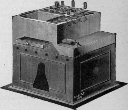
Fig. 31. Paraffin-embedding Oven.
After dehydration the specimen is placed in a clearing agent - chloroform or xylol - both of which are suitable when embedding in paraffin. The clearing agents replace the alcohol in the cells, and at the same time render the tissues transparent. From the clearing agent the specimen is placed in a weak solution of paraffin, dissolved xylol, or chloroform. The strength of the paraffin solution is gradually increased until it consists of pure paraffin. The temperature of the paraffin-embedding oven (Fig. 31) should not be much higher than the melting-point of the paraffin.
The specimen is now ready to be embedded. First make a mould of cardboard or a lead-embedding frame (Fig. 32), melt the paraffin, and then place the specimen in a manner that will facilitate cutting. Remove the excess of paraffin and cut when desired.
In using the collodion method for embedding fibrous specimens, as wood, bark, roots, etc., the specimen is first fixed with picric acid, washed with water, cleared in ether-alcohol, embedded sucessively collected five and twelve per cent ether-alcohol collodion solution, and finally embedded in a pure collodion bath.
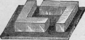
Fig. 32. Paraffin Blocks.
Continue to:
My Books

