Specific Techniques For Care Of Patients With Ileal Bladder (Artificial Bladder)
Description
This section is from the book "Cancer Manual For Public Health Nurses", by National Cancer Institute. Also available from Amazon: Cancer Nursing: A Manual For Public Health Nurses.
Specific Techniques For Care Of Patients With Ileal Bladder (Artificial Bladder)
1. Equipment-A variety of rubber and disposable plastic bags are available for artificial bladder care. They are all similar in principle in that they can be attached to the abdominal wall by means of a collar which fits over the stoma (see diagram, page 60). The opening in the collar of some bags must be individually cut to fit the outer circumference of the stoma. Other collars come with openings of specified size. The collar is attached to the skin by means of liquid adhesive applied with a brush or applicator. Some of the newer collection bags have discs which are already coated with the necessary adhesive. See procedure for changing ileal bladder bags. Equipment needed to apply a bag includes:
(1) soap and water (or a substitute) to cleanse the skin
(2) skin preparation to protect the skin-tincture of benzoin or any substitute ordered by the physician
(3) cleaning brush (for bag)
(4) belt for holding the bag in place
(5) solution for cleaning bag-soap and water with a table-spoonful of vinegar or a pint of water containing, 1 teaspoon of clorox and one-half cup of vinegar
(6) liquid adhesive, if needed
The directions given apply only to the use of ileal bladder bags
ILEAL BLADDER. Diagram D


Diagram E with double faced adhesive discs. Be sure to follow specific directions for applying other types of bags. (Figure 13).
3. Care of the Bag:
(1) syringe or rinse with plain cool tap water to remove mucus
(2) wash with bland soap and water to which vinegar or clorox has been added
(3) rinse thoroughly and let dry (never over heat); separate sides of bag to facilitate drying
4. General Information
(1) It is recommended that at least two bags be used. This provides time for the bags to air and minimizes the odor.
(2) Bags should be changed as often as needed.
It would be impossible to enumerate all of the problems which
?. Procedure (or Changing Ileal Bladder Rag

1. Empty bag of urinary contents through opening at bottom of bag.
2. Remove belt.

3. Gently peel bag from abdomen
4. Cleanse skin around bladder opening. Let dry.
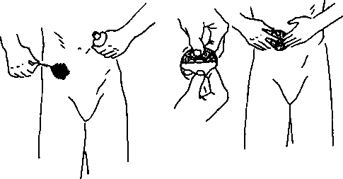
5. Paint area around artificial opening with tincture of benzoin using cotton applicator. Let dry for one minute.
6. Remove protective covering carefully from adhesive disc. 7. Center adhesive disc over artificial bladder opening and press firmly to skin area painted with benzoin.

8. Carefully remove outer protective covering from disc.

9. Center bag and quickly press into place over artificial bladder opening.
10. Attach belt to plastic frame and fasten firmly for watertight seal.
Compound tincture of benzoin is essential for proper application when double - faced adhesive discs are used.
Figure 13 patients with radical surgery of this nature may face. The adjustment varies markedly, but most patients accept the problems which accompany the procedure and appreciate the added time and comfort which they are given. In many instances the woman is very grateful for the relief from pain which the surgery affords. However, most of these patients need a tremendous amount of support from everyone concerned. The nurse is in a unique position to offer constructive help in assisting the patient to solve her problems about personal grooming, control of odors, or ways of expediting the time-consuming procedures which must be performed. She can make practical suggestions relative to what can be done when equipment does not "work well," when leakage occurs, bags do not stay cemented to the skin, etc. She may also help the patient accept the changes which have been made in her anatomy-for example, the feeling of lightness in the pelvic basin due to the extensive resection which has been performed. She may also be supportive in furthering the many psychological adjustments which must inevitably be made. Patients often express worries about the possibility of odors and accidents involving spillage of urine and/or feces. Others worry about the fit of their clothes over the collection bags. Many patients fear rejection by other family members, especially those closest to them. There is sometimes a tendency to withdraw from other people.
Obviously there will be some problems which the nurse will not be able to help the patient solve. However, she can serve a useful function in her role as a listener. In addition she can often refer the patient to the community resources which are most suited to meeting the needs of the patient or the family.
Radiation Therapy
Before discussing radiation as a form of therapy it might be helpful to review a few simple facts about radiation, the interaction of matter, and the various forms of ionizing radiation used in therapy.
An element is a substance such as hydrogen, carbon or uranium which cannot be broken down into another substance by chemical methods. Combination of elements are known as chemical compounds and these can be separated rather easily into their constituent parts.
The smallest unit of a compound is a molecule and this is composed of atoms which are chemically bound together. The number of atoms in a molecule varies with the compound. An atom can be defined as the simplest unit into which an element can be divided and still retain the chemical properties of the original element.
Certain naturally occurring elements such as radium or uranium are unstable, and give off ionizing rays which constantly fly off into space. These rays cannot be seen, but they are capable of penetrating anything in their path to varying degrees. There are three of these rays with which we are concerned: Alpha, beta, and gamma (see glossary).
It should be remembered that radium is a naturally occurring radioisotope. In addition to the naturally occurring radioisotopes, we now have hundreds of artificially produced radioisotopes which are prepared in special installations like the reactor at Oak Ridge, Tenn. Radioisotopes may be purchased from either Oak Ridge or some of the drug companies. These radioisotopes are widely used in hospitals and clinics for both diagnosis and treatment.
X-Ray And Teletherapy
X-rays are similar to gamma rays and achieve much the same effect when used therapeutically.
The radiologist who employs X-ray with the intent to effect a cure has a wide range of treatment possibilities from which to choose. These vary from conventional voltage X-ray therapy of 200-250 kilovolts to supervoltage X-ray ranging from 500 to 2,000 kilovolts. In addition to the standard X-ray unit which is familiar to almost everyone, there are now many teletherapy units in hospitals and clinics throughout the country. These look similar to X-ray units but unlike the X-ray apparatus the source of power is not electricity. Instead the machine houses a radioactive substance (a radioisotope) such as cobalt, iridium, or cesium. These radioisotopes emit gamma rays and these rays have the same therapeutic effect as X-rays. Many radiologists feel that this method of treatment causes less damage to the skin.
In addition to the above methods of treatment a technique called rotational X-ray therapy or moving beam technique is sometimes employed. The patient may be rotated in the path of a fixed beam, or the X-ray beam may be rotated while the patient remains stationary.
Continue to:
My Books

