The Epidermis And Periderm. Part 5
Description
This section is from the "Histology of Medicinal Plants" book, by William Mansfield. Also see Amazon: Histology of Medicinal Plants.
The Epidermis And Periderm. Part 5
Periderm
The periderm is the outer protective covering of the stems and roots of mature shrubs and trees. The periderm replaces the epidermis. The periderm may be composed of cork cells, stone cell-cork, or a mixture of cork, parenchyma, nbres, stone cells, etc.
Cork Periderm
The typical periderm is made up of cork cells. Cork cells vary in appearance, according to the part of the cell viewed.
On surface view (Plate 16, Fig. A) the cork cells are angled in outline and are made up of from four to seven side walls; five- and six-sided cells are more common than the four- and seven-sided cells. Surface sections of cork cells show their length and width. These side walls usually appear nearly white, while the end wall, particularly of the outermost cork cells, usually appears brown or reddish-brown, or in some cases nearly black.


Plate 14. Multicellular Multiseriate Branched Hairs.
1. Apical hairs arnica (Arnica montana, L.).
2. Basal hairs arnica (Arnica montana, L.).
3. Apical hairs grindelia (Grindelia squarrosa, [Pursh] Dunal).
4. Basal hairs grindelia (Grindelia squarrosa, [Pursh] Dunal).

Plate 15. Multicellular Multiseriate Branched Hairs.
1. Apical hairs boneset {Eupatorium perfoliatum, L.).
2. Basal hairs boneset (Eupatorium perfoliatum, L.).
3. Apical hairs life-everlasting (Gnaphalium obtusifolium, L.).
4. Basal hairs life-everlasting (Gnaphalium obtusifolium, L.).
Cork cells on cross-section are rectangular in form, and they are arranged in superimposed rows, the number of rows being gradually increased as the plant grows older. Such an increase in the number of rows of cork cells is shown in the cross-section of cascara sagrada (Plate 16, Fig. C).
Cork cells fit together so closely that there is no intercellular spaces between the cells. In this case two rows of cork cells occupy no greater space than the solitary row of cork cells immediately over and external to them. As a rule, the outermost layers of cork cells have a narrower radial diameter than the cork cells of the underlying layers. This is due to the fact that these outer cells are stretched as the stem increases in diameter. This view shows the height of cork cells, but not always the length, which will, of course, vary according to the part of the cell cut across. In a section a few millimeters in diameter, however, all the variations in size may be observed. The color of the walls is nearly white.
The cavity may contain tannin or other substances. When tannin is present, the cavity is of a brownish or brownish-red color, or it may be nearly black. Most barks appear devoid of any colored or colorless cell contents.
The radial section (Plate 16, Fig. B) of cork cells shows the height of the cells and the width of the cells at the point cut across. Some cells will be cut across their longest diameter, while others will be cut across their shortest diameter. Cork cells are, therefore, smaller in radial section than they are in cross-section. The color of the walls is white, and the color and nature of the cell contents vary for the same reasons that they vary in cross-sections.
The number of layers of cork cells occurring in cross- and radial-sections varies according to the age of the plant, to the type of plant, and to the conditions under which the plant is growing.
The number of layers of cork cells is not of diagnostic importance, nor is the surface view of cork cells diagnostic except in certain isolated cases.

Plate 16. Periderm of Cascara Sagrada (Rhamnus purshiana, D.C.).
A. I, Outline of cork cells; 2, Line of contact of adjoining cork cells.
B. Radial longitudinal section of cascara sagrada. 1, Cork cells; 2,Phel-logen; 3, Forming parenchyma cells; 4, Cortical parenchyma cells.
C. Cross-section of cascara sagrada. I, Cork cells; 2, Phellogen; 3, Forming parenchyma cells; 4, Cortical parenchyma cells.
The presence or absence of cork or epidermal tissue in powders must always be noted. The presence of cork enables one to distinguish Spanish from Russian licorice. In like manner, the presence of epidermis enables one to distinguish the pharma-copoeial from the unofficial peeled calamus. The absence of epidermis in Jamaica ginger is one of the means by which this variety is distinguished from the other varieties of ginger, etc.
In canella alba the periderm is replaced by stone cell-cork. That is, the cells forming the periderm are of a typical cork shape, but the walls are lignified, unequally thickened, and the inner or thicker walls are strongly porous, and the walls are of a yellowish color. Stone cell-cork forms the periderm of clove bark also, but the cells are narrower and longer, and the inner wall is not so thick or porous as is the case in canella alba bark.
Stone Cell Periderm
In canella alba (Plate 17, Fig. B) cork periderm is frequently replaced by stone cells, particularly in the older barks. These stone cells form the periderm because they replace the cork periderm, which fissures and scales off as the root increases in diameter.
The side and end walls of cork cells are of nearly uniform diameter. Exceptions occur, but they are not common. In buchu stem (Plate 101, Fig. 3), the cork cells have thick outer walls, but thin sides and inner walls. The cell cavity contains reddish-brown deposits of tannin.
Parenchyma And Stone Cell Periderm
As the trees and shrubs increase in diameter, cracks or fissures occur in the periderm, or corky layer. In such cases the phellogen cells divide and redivide in such manner as to cut off a portion of the parenchyma cells, stone cells, and fibres of the cortex which is inside of and below the fissure. All the parenchyma cells, etc., exterior to the newly formed cork cells soon lose their living-cell contents, since their food-supply is cut off by the impervious walls of the cork cells. In time they are forced outward by the developing cork cells until they partially or completely fill the break in the periderm. In white oak bark (Plate 18), as in other barks, a large part of the periderm is composed of dead and discolored cortical cells.
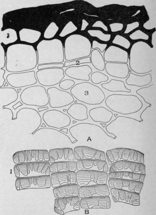
Plate 17. A. Cross-section of Mandrake Rhizome (Podophyllum peltatum, L.).
1. Epidermis.
2. Phellogen.
3. Cortical parenchyma.
B. Stone cell periderm of white cinnamon (Canella alba, Murr.).

Plate 18. Periderm of White Oak (Quercus alba, L.).
I. Outer layer of cork cells. 2. Cortical parenchyma cells. 3. Stone cells. 4. Phellogen. 5. Cortical parenchyma cells.
Origin Of Cork Cells
The cork cells are formed by the meristimatic phellogen cells, which originate from cortical parenchyma. These cells divide into two cells, the outer changing into a cork cell, while the inner cell remains meristimatic. In other instances the outer cell remains meristimatic, while the inner cell changes into a cortical parenchyma cell. The development of a cortical parenchyma cell from a divided phellogen cell is shown in Plate 101, Fig. 6. Both the primary and secondary cork cells originate from the phellogen or cork cambrium layer. Cork cells do not contain living-cell contents; in fact, in the majority of medicinal barks the cork cells contain only air.
The walls of typical cork cells are composed, at least in part, of suberin, a substance which is impervious to water and gases. In certain cases layers of cellulose, lignin, and suberin have been identified. Suberin, however, is present in all cork cells, and in some cases all of the walls of cork cells are composed of suberin.
Suberized cork cells are colored yellow with strong sodium hydroxide solutions and by chlorzinciodide.
Continue to:
My Books

