Chapter XXVII. Diseases Of The Brain. The Dura Mater
Description
This section is from the book "A Manual Of Pathology", by Guthrie McConnell. Also available from Amazon: A Manual Of Pathology.
Chapter XXVII. Diseases Of The Brain. The Dura Mater
Hyperemia may be active as a result of injuries or disease of the skull. Passive hyperemia may follow thrombosis of the venous sinuses. Neither of the above can be well recognized post-mortem, as by that time hypostasis has taken place and the blood has sought the lower levels.
Thrombosis of the sinuses is frequently secondary to extension of inflammation from adjacent bony structures, as in mastoid and middle-ear disease. It also occurs in infectious diseases, and is usually located in the superior longitudinal sinus. Cerebral softening or abscess formation with pulmonary or cardiac embolism may follow thrombosis and cause death.
Hemorrhage is commonly due to injury, and may take place on the internal or external surface of the dura. A comparatively large amount of blood may collect between the skull and the dura - an internal cephalhematoma - and give rise to serious compression symptoms. Small hemorrhages, frequently multiple, may be found in the substance of the dura after death by suffocation.
Inflammation Of The Dura
Acute pachymeningitis is the result of infection following injury or disease of the skull. It may be local or general, and is characterized by the presence of pus. The dura is much thickened and swollen by round-cell infiltration, and is covered by a layer of purulent material. Hemorrhagic pachymeningitis is found in the old, the insane, and in alcoholics, usually in the area that is supplied by the middle meningeal artery. It consists of a chronic inflammation of the internal layer of the dura mater, characterized by the formation of layers of new, delicate connective tissue with numerous very thin-walled blood-vessels from which the blood is prone to escape. At times this may be so extensive as to simulate a hemorrhage. The coloring-matter may be absorbed and leave a collection of serous fluid - a hygroma.
In more advanced stages the new membrane may become greatly thickened, its outermost layers being changed into dense fibrous tissue with the obliteration of the vessels.
Chronic internal pachymeningitis is of obscure etiology, probably hematogenic, and is usually accompanied by disease of the pia and arachnoid. It is characterized by the deposition of numerous layers of fibrinous exudation upon the internal surface of the dura. These gradually undergo fibrous replacement, with frequently the formation of many new capillaries. The dura becomes more adherent to the bone, and calcareous infiltration is sometimes encountered.
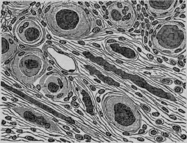
Fig. 182. - Section of a Psammoma of the Dura Mater. X 200 (Ziegler).
Tuberculosis of the dura usually follows tuberculous disease of' the bones of the skull or of the pia-arachnoid. It may be present as miliary tubercles or as large caseous masses.
Syphilis may give rise to a pachymeningitis fibrosa, causing a dense thickening of the dura. It may also be present as gummata, which may have originated either within the dura or within the bones of the skull, and have secondarily invaded the membrane.
Tumors
The most common is the sarcoma, which maybe either spindle or round-celled, and quite often alveolar. These growths extend from the inner surface of the dura toward the brain. They may be flat or more elevated, and vary greatly in size. If they form on the outer surface of the dura, they may cause absorption of the bone and perforation. If the blood-supply is very rich, these growths are called angiosarcoma. Endothelioma may develop upon the inner surface of the membrane. Other forms of primary tumors are rare, psammoma, lipoma, and fibroma seldom occurring.
Secondary tumors may follow malignant disease of neighboring structures - may be sarcoma, glioma, or carcinoma.
Parasites are rare; the echinococcus and the Cysticercus cellulosoe have been described.
Continue to:
My Books

