Roots And Rhizomes. Part 2
Description
This section is from the "Histology of Medicinal Plants" book, by William Mansfield. Also see Amazon: Histology of Medicinal Plants.
Roots And Rhizomes. Part 2
Cross-Section Spigelia Rhizome
The cross-section of spigelia rhizome (Plate 91) is as follows:
Epidermis
The epidermal cells are nearly angled and free of cell contents.
Cortex
The cortical parenchyma cells are usually slightly tangentially elongated. The cells of the outer layers are larger than the cells of the inner layers.
Phloem
The phloem contains sieve cells and phloem parenchyma. The sieve cells are small, angled cells with thin, white walls.
The phloem parenchyma cells resemble the sieve cells, but they are larger.
Cambium
The cambium cells are rectangular, and they are usually not clearly seen because the walls are partially collapsed.
Xylem
The xylem is composed of vessels, wood parenchyma, medullary rays, and pith parenchyma.
Vessels
The vessels are slightly angled in outline and few in number.
Wood Parenchyma
The wood parenchyma cells are small and angled.
Medullary Rays
The medullary ray cells are tangentially elongated, but in structure resemble the wood parenchyma cells.
Pith Parenchyma
The pith parenchyma cells are rounded in outline and contain small, simple, rounded starch grains.
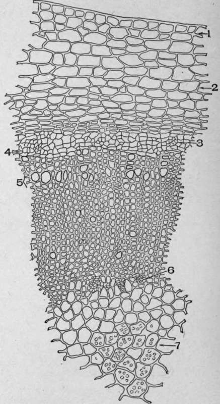
Plate 91. Cross-Section of Rhizome of Spigelia marylandica, L.
I. Epidermis. 2. Cortical parenchyma. 3. Phloem. 4. Cambium.
5. Xylem. 6. Internal phloem. 7. Pith with starch.
Cross-Section Ruellia Rhizome
The cross-section of ruellia rhizome (Plate 92) differs from the structure of spigelia rhizome, It is as follows:
Epidermis
The epidermal cells vary in shape from nearly square to oblong, and they are rilled with dark-brown cell contents.
Cortex
The cortex contains parenchyma and stone cells.
The outer layer of the cortical parenchyma cells are variable in size and many of the cells contain deposits of calcium carbonate and dark cell contents; the inner parenchyma cells are larger and they are free of the dark-brown cell contents, but many of the cells contain deposits of calcium carbonate.
Stone cells with thick, white, porous, and striated walls occur in among the cortical parenchyma cells.
Phloem
The phloem contains sieve cells, phloem, parenchyma, and bast fibres.
The sieve cells are small and with thin, white, angled walls.
The phloem parenchyma cells resemble the sieve cells, but they are larger.
The bast fibres occur singly or in groups of two or three. The walls are white, non-porous, and non-striated.
Cambium
The cambium layer is composed of rectangularly shaped cells, which are frequently obliterated.
Xylem
The xylem contains vessels, wood parenchyma, and medullary rays.
The vessels are large, rounded cells with thick walls.
The wood parenchyma consists of thick-walled cells of irregular size and form.
The medullary rays are tangentially elongated and rectangular in form.
Pith Parenchyma
The pith parenchyma cells are rounded in outline and as large as the cortical parenchyma cells.

Plate 92. Cross-Section of Rhizome of Ruellia ciliosa, Pursh.
1. Epidermis. 2. Cystolith. 3. Stone cell. 4. Cortical parenchyma. 5, Bast fibres. 6. Pericycle. 7. Xylem. 8. Pith.
Powdered Pink Root
When the roots and rhizomes of spigelia are powdered (Plate 93) they show the following structure:
The epidermal cells are small and brownish on surface view, varying in size from 13 by 18 micromillimeters to 31 by 40 micromillimeters. When associated with parenchyma they appear as black masses. The cortical parenchyma cells are rounded and vary in size from 23 by 26 micromillimeters to 37.5 by 90 micromillimeters. Many of the cells from the root contain larger quantities of minute single rounded starch grains varying in size from 1 micromillimeter to 4 micromillimeters. The larger round single starch grains are found in both the cortical and pith parenchyma of the rhizome. They vary in size from 5 micromillimeters to 18 micromillimeters. The conducting elements are pitted tracheids varying from 10 micromillimeters to 38 micromillimeters in diameter. A few pitted and annular vessels are also found. The only fibres occurring are found in the xylem. They are not a prominent feature of the powder, as their walls break up into minute fragments. The pith parenchyma varies in size from 13 by 19 micromillimeters to 75 by 82.5 micromillimeters. It is in these cells that the largest starch grains occur.
Distinguishing Diagnostic Characters Of The Powder
1. Parenchyma with starch.
2. Dark masses of epidermal tissue.
3. Spigelia should contain starch, and it should not contain cystoliths, stone cells, or long, white-walled bast fibres.
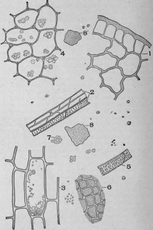
Plate 93. Powdered Spigelia marylandica, L.
1. Epidermis and cortical parenchyma. 2. Tracheids and fibres. 3. Parenchyma cells of the root containing the small starch grains, longitudinal view. 4. Parenchyma of the rhizome containing the large starch grains, transverse view. 5. Tracheids. 6. Surface view of the epidermal cells. 7. Starch scattered through the field. 8 and 8'. Dark masses of epidermal and underlying tissue.
Continue to:
My Books

