Roots And Rhizomes. Part 3
Description
This section is from the "Histology of Medicinal Plants" book, by William Mansfield. Also see Amazon: Histology of Medicinal Plants.
Roots And Rhizomes. Part 3
Powdered Ruellia Root
When the roots of ruellia root and rhizome are powdered (Plate 94) they show the following structure:
The epidermal cells vary from 7.8 by 15.6 micromillimeters to 15.1 by 16.6 micromillimeters. The cell contents are dark and the walls are light. A few rows of the outer cortical parenchyma cells of both the rhizome and the root have dark cell contents and white walls. The dark contents disappear toward the phloem. The cortical cells vary from 13.6 by 14.3 micro-millimeters to 89.5 by 90.9 micromillimeters. In the cortical parenchyma cells of the rhizome are found the short, broad cystoliths measuring up to 52 by 62 micromillimeters. In the corresponding cells of the root are found the long, narrow cystoliths which measure up to 68.4 by 187.2 micromillimeters. Scattered throughout the powder are seen three distinct types of sclerids (stone cells) which are associated with the cortical parenchyma of both the stem and the root. Most of them are found, however, in the roots. First, the short, broad stone cells from the stem basis have square ends; the walls vary from 13 to 19.5 micromillimeters in thickness with branching pores which extend toward the adjacent cell. These sclerids vary in size from 52 by 54.6 micromillimeters to 45 by 130 micromillimeters. Secondly, the long stone cells from the root vary from 32 by 96 micromillimeters to 45.5 by 542.5 micromillimeters with walls 16 micromillimeters thick. The width of the cell and the thickness of the wall vary but little throughout their entire length. The third type of stone cell also from the root has unequally thickened walls and the ends are square or blunt. A few long, narrow, colorless, thin-walled bast fibres also occur. They are 13 micromillimeters wide, with walls 3.9 micromillimeters thick. Annular spiral and pitted vessels are also found scattered throughout the powder.
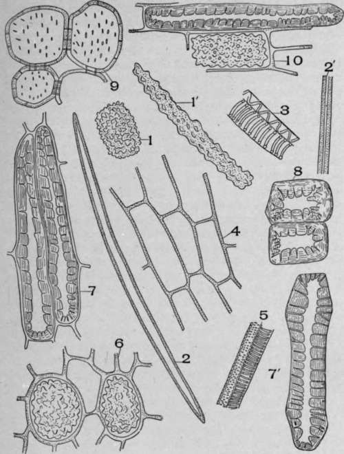
Plate 94. Powdered Ruellia ciliosa, Pursh.
I. Short, broad cystoliths from the rhizome, 1'. Long cystoliths from the root. 2 and 2'. Long, narrow, white-walled bast fibres. 3. Tracheal tissue from the xylem of the stem. 4. Root parenchyma. 5. Tracheal tissue from the xylem of the root. 6. Cortical parenchyma cells from the rhizome with short, broad cystoliths. 7 and 7'. Long, thick-walled sclerids from the root. 8. Short, broad sclerids from the stem. 9. Pitted pith parenchyma from the stem with intercellular space. 10. Parenchyma of the root with sclerid and cystolith, longitudinal view.
The diagnostic characters of the powder are:
1. The short, broad, and long, narrow cystoliths.
2. The short, broad, and long, narrow sclerids.
3. The long, narrow, thin, white-walled bast fibres.
In poke root, ipecac, sarsaparilla, and veratrum are raphides. In belladonna and horse-nettle roots are micro-crystals. In calumba, stillingea, krameria, licorice, scamony root are prisms. In saponaria, jalap, althea, spikenard, rumex, rhubarb are rosette crystals. In pleurisy roots both prisms and rosettes occur.
In gentian, senega, symphytuns, lovage, parsley, inula, echinacea, angelica, burdock, and chicory no crystals of any-kind occur. Root hairs occur in cross-sections of sarsaparilla root and false unicorn, but with these exceptions: root hairs do not occur on roots, because the younger part of the root with root hairs is not removed from the soil when the drug is collected. In sarsaparilla root there are several layers of hypodermal cells; in most roots there are no hypodermal cells. In the non-woody roots or the roots of herbs the parenchyma cells form the greater part of the tissues of the root. In ruellia root are stone cells; in spigelia root and many other roots there are no stone cells. In ruellia root are bast fibres; in spigelia, gentian, ipecac, chicory, dandelion, Symphytum, and lovage no bast fibres occur. In all the woody roots there is a periderm consisting of typical cork cells, as in black haw; or stone cells, as in asclepias; or of a mixture of lifeless parenchyma, medullary rays, etc., as in Oregon grape root.
Woody roots have a phellogen layer which is absent in the non-woody roots.
The numbers of layers of cortical parenchyma differ in the same root according to its age, but for a given root there is a normal variation.
The number of layers of cortical parenchyma in proportion to other cells is less in woody roots.
In woody roots there is no endodermis. The cambium in these cases shows clearly between the phloem and the xylem part of the fibro-vascular bundle.
In woody roots the wood fibres are well developed and form a large part of the root, and the medullary rays have pitted side and end walls.
The description given above of ruellia root is not typical of all roots, but the structure represents the greater number of the elements that it is possible to find in a root. In many roots, for instance, there are no stone cells, in others no epidermis and no endodermis. In asclepias, aconite, and calumba stone cells occur. In Symphytum, chicory, dandelion, burdock, elecampane, Pyrethrum, gentian, and senega no stone cells occur. In aconite, althea, asclepias, belladonna, bryonia, columba, ipecac, jalap, krameria, sarsaparilla, scamony, stillingea, and rumex are characteristic starch grains. Symphytum, chicory, dandelion, burdock, elecampane, and Pyrethrum contain inulin, but no starch. In saponaria, gentian, and senega neither starch nor inulin occurs.
When studying roots the nature of the epidermis or the periderm must be considered, as also the number of layers of cortical parenchyma; the occurrence, distribution, and amount of stone cells when present; the presence or absence of the endodermis; the occurrence and structure of bast fibres when present; the nature of the cambium cells; the width and structure of the medullary rays, the size of the wood fibres and wood parenchyma, and the nature of the cell contents and the arrangement of the fibro-vascular bundle.
Continue to:
My Books

